Explore
 Validate
 Learn
Learn Western blot
 ELISA
ELISAAntibody data
- Antibody Data
- Antigen structure
- References [0]
- Comments [0]
- Validations
- Western blot [5]
- Immunocytochemistry [2]
- Immunohistochemistry [1]
Submit
Validation data
Reference
Comment
Report error
- Product number
- GTX112677 - Provider product page

- Provider
- GeneTex
- Proper citation
- GeneTex Cat#GTX112677, RRID:AB_10620428
- Product name
- APP antibody
- Antibody type
- Polyclonal
- Reactivity
- Human, Mouse, Rat
- Host
- Rabbit
No comments: Submit comment
Supportive validation
- Submitted by
- GeneTex (provider)
- Main image
- Experimental details
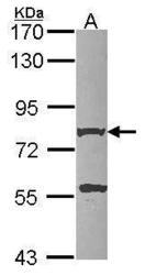
- Sample (30 ug of whole cell lysate) A: HCT116 7.5% SDS PAGE amyloid beta A4 antibody GTX112677 diluted at 1:1000
- Validation comment
- WB

- Submitted by
- GeneTex (provider)
- Main image
- Experimental details
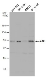
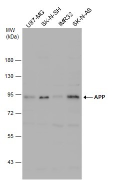
- Various whole cell extracts (30 £gg) were separated by 7.5% SDS-PAGE, and the membrane was blotted with APP antibody (GTX112677) diluted at 1:500.
- Submitted by
- GeneTex (provider)
- Main image
- Experimental details
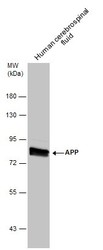
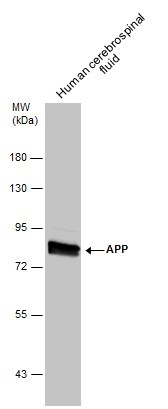
- Human tissue extract (30 £gg) was separated by 7.5% SDS-PAGE, and the membrane was blotted with APP antibody (GTX112677) diluted at 1:500.
- Submitted by
- GeneTex (provider)
- Main image
- Experimental details
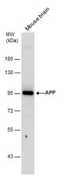
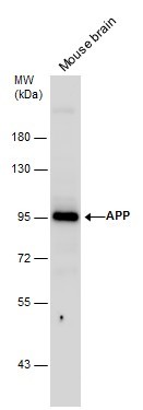
- Mouse tissue extract (50 £gg) was separated by 7.5% SDS-PAGE, and the membrane was blotted with APP antibody (GTX112677) diluted at 1:500.
- Submitted by
- GeneTex (provider)
- Main image
- Experimental details
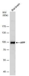
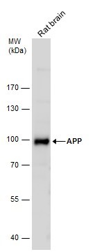
- Rat tissue extract (50 £gg) was separated by 7.5% SDS-PAGE, and the membrane was blotted with APP antibody (GTX112677) diluted at 1:500.
Supportive validation
- Submitted by
- GeneTex (provider)
- Main image
- Experimental details
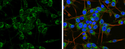
- APP antibody detects APP protein at cytoplasm by immunofluorescent analysis.Sample: U-87 MG cells were fixed in 4% paraformaldehyde at RT for 15 min.Green: APP protein stained by APP antibody (GTX112677) diluted at 1:100.Blue: Hoechst 33342 staining.

- Submitted by
- GeneTex (provider)
- Main image
- Experimental details
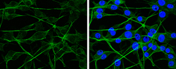
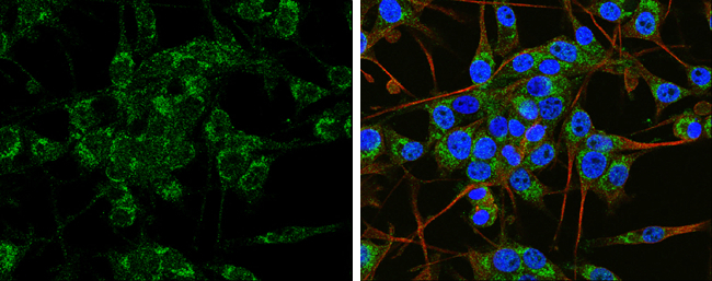
- APP antibody detects APP protein at cytoplasm by immunofluorescent analysis.Sample: SH-SY5Y cells were fixed in 4% paraformaldehyde at RT for 15 min.Green: APP protein stained by APP antibody (GTX112677) diluted at 1:100.Red: beta Tubulin 3/ TUJ1 protein stained by beta Tubulin 3/ TUJ1 antibody (GTX631836) diluted at 1:200.Blue: Hoechst 33342 staining.
Supportive validation
- Submitted by
- GeneTex (provider)
- Main image
- Experimental details
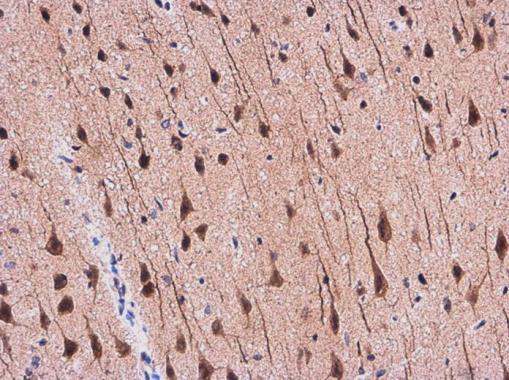
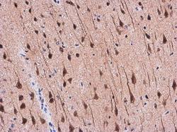
- APP antibody detects APP protein at cytoplasm in Rat brain by immunohistochemical analysis. Sample: Paraffin-embedded Rat brain. APP antibody (GTX112677) diluted at 1:500.