Explore
 Validate
 Learn
Learn Western blot
 Immunohistochemistry
Antibody data
- Antibody Data
- Antigen structure
- References [8]
- Comments [0]
- Validations
- Western blot [1]
- Immunocytochemistry [2]
- Other assay [4]
Submit
Validation data
Reference
Comment
Report error
- Product number
- 14-9749-82 - Provider product page

- Provider
- Invitrogen Antibodies
- Product name
- APP (Amyloid Precursor Protein) Monoclonal Antibody (22C11), eBioscience™
- Antibody type
- Monoclonal
- Antigen
- Other
- Description
- Description: The monoclonal antibody 22C11 recognizes human, mouse, and rat APP (Amyloid Precursor Protein). APP is expressed in high abundance in the central nervous system and has three major isoforms resulting from alternative splicing. APP plays a role in synaptic formation and repair, anterograde neuronal transport, iron export, and hormonal regulation. Secreted APP (sAPP) may have neuroprotective effects against neurotoxic insult, oxidative stress, and excitotoxicity. APP belongs to a family that contains at least two homologs, amyloid precursor-like proteins 1 and 2 (APLP1 and APLP2). Similarities between APP and APLP, especially APLP2, suggest that APLP could share and compensate for the function of APP. Proteolytic cleavage of APP results in the generation of beta amyloid, which is the primary component of senile plaques. Senile plaques are one of the major histopathologic features of Alzheimer's disease. Abnormal regulation and processing of APP also plays a role in Down's syndrome, early onset familial Alzheimer's disease, cerebral hemorrhage, and arthritis. This 22C11 antibody reacts with pre-A4 and recognizes all three isoforms of APP (immature, sAPP, and mature). This 22C11 antibody is known to cross react with APLP2. Applications Reported: This 22C11 antibody has been reported for use in microscopy, and immunocytochemistry. Applications Tested: This 22C11 antibody has been tested by immunocytochemistry of methanol-fixed cells and can be used at less than or equal to 2.5 µg/mL. It is recommended that the antibody be carefully titrated for optimal performance in the assay of interest. Purity: Greater than 90%, as determined by SDS-PAGE. Aggregation: Less than 10%, as determined by HPLC. Filtration: 0.2 µm post-manufacturing filtered.
- Reactivity
- Human, Mouse, Rat
- Host
- Mouse
- Isotype
- IgG
- Antibody clone number
- 22C11
- Vial size
- 100 µg
- Concentration
- 0.5 mg/mL
- Storage
- 4° C
Submitted references Melanoma-Secreted Amyloid Beta Suppresses Neuroinflammation and Promotes Brain Metastasis.
microRNA-425 loss mediates amyloid plaque microenvironment heterogeneity and promotes neurodegenerative pathologies.
GSAP regulates lipid homeostasis and mitochondrial function associated with Alzheimer's disease.
APOE4 exacerbates synapse loss and neurodegeneration in Alzheimer's disease patient iPSC-derived cerebral organoids.
Depletion of the AD Risk Gene SORL1 Selectively Impairs Neuronal Endosomal Traffic Independent of Amyloidogenic APP Processing.
Survival of cultured neurons from amyloid precursor protein knock-out mice against Alzheimer's amyloid-beta toxicity and oxidative stress.
Amyloid-like properties of peptides flanking the epitope of amyloid precursor protein-specific monoclonal antibody 22C11.
Subcellular localization of amyloid precursor protein in senile plaques of Alzheimer's disease.
Kleffman K, Levinson G, Rose IVL, Blumenberg LM, Shadaloey SAA, Dhabaria A, Wong E, Galán-Echevarría F, Karz A, Argibay D, Von Itter R, Floristán A, Baptiste G, Eskow NM, Tranos JA, Chen J, Vega Y Saenz de Miera EC, Call M, Rogers R, Jour G, Wadghiri YZ, Osman I, Li YM, Mathews P, DeMattos RB, Ueberheide B, Ruggles KV, Liddelow SA, Schneider RJ, Hernando E
Cancer discovery 2022 May 2;12(5):1314-1335
Cancer discovery 2022 May 2;12(5):1314-1335
microRNA-425 loss mediates amyloid plaque microenvironment heterogeneity and promotes neurodegenerative pathologies.
Hu YB, Zhang YF, Ren RJ, Dammer EB, Xie XY, Chen SW, Huang Q, Huang WY, Zhang R, Chen HZ, Wang H, Wang G
Aging cell 2021 Oct;20(10):e13454
Aging cell 2021 Oct;20(10):e13454
GSAP regulates lipid homeostasis and mitochondrial function associated with Alzheimer's disease.
Xu P, Chang JC, Zhou X, Wang W, Bamkole M, Wong E, Bettayeb K, Jiang LL, Huang T, Luo W, Xu H, Nairn AC, Flajolet M, Ip NY, Li YM, Greengard P
The Journal of experimental medicine 2021 Aug 2;218(8)
The Journal of experimental medicine 2021 Aug 2;218(8)
APOE4 exacerbates synapse loss and neurodegeneration in Alzheimer's disease patient iPSC-derived cerebral organoids.
Zhao J, Fu Y, Yamazaki Y, Ren Y, Davis MD, Liu CC, Lu W, Wang X, Chen K, Cherukuri Y, Jia L, Martens YA, Job L, Shue F, Nguyen TT, Younkin SG, Graff-Radford NR, Wszolek ZK, Brafman DA, Asmann YW, Ertekin-Taner N, Kanekiyo T, Bu G
Nature communications 2020 Nov 2;11(1):5540
Nature communications 2020 Nov 2;11(1):5540
Depletion of the AD Risk Gene SORL1 Selectively Impairs Neuronal Endosomal Traffic Independent of Amyloidogenic APP Processing.
Knupp A, Mishra S, Martinez R, Braggin JE, Szabo M, Kinoshita C, Hailey DW, Small SA, Jayadev S, Young JE
Cell reports 2020 Jun 2;31(9):107719
Cell reports 2020 Jun 2;31(9):107719
Survival of cultured neurons from amyloid precursor protein knock-out mice against Alzheimer's amyloid-beta toxicity and oxidative stress.
White AR, Zheng H, Galatis D, Maher F, Hesse L, Multhaup G, Beyreuther K, Masters CL, Cappai R
The Journal of neuroscience : the official journal of the Society for Neuroscience 1998 Aug 15;18(16):6207-17
The Journal of neuroscience : the official journal of the Society for Neuroscience 1998 Aug 15;18(16):6207-17
Amyloid-like properties of peptides flanking the epitope of amyloid precursor protein-specific monoclonal antibody 22C11.
Hilbich C, Mönning U, Grund C, Masters CL, Beyreuther K
The Journal of biological chemistry 1993 Dec 15;268(35):26571-7
The Journal of biological chemistry 1993 Dec 15;268(35):26571-7
Subcellular localization of amyloid precursor protein in senile plaques of Alzheimer's disease.
Kawai M, Cras P, Richey P, Tabaton M, Lowery DE, Gonzalez-DeWhitt PA, Greenberg BD, Gambetti P, Perry G
The American journal of pathology 1992 Apr;140(4):947-58
The American journal of pathology 1992 Apr;140(4):947-58
No comments: Submit comment
Supportive validation
- Submitted by
- Invitrogen Antibodies (provider)
- Main image

- Experimental details
- Western blot was performed using Anti-APP (Amyloid Precursor Protein) Monoclonal Antibody (22C11), eBioscience™(Product # 14-9749-80) and a 120, 90 kDa band corresponding to APP (Amyloid Precursor Protein) was observed in Mouse Brain and Rat Brain as compared to Mouse and Rat Skeletal Muscle which are reported to be negative. Tissue extracts (30 µg lysate) of Mouse Brain (Lane 1), Mouse Skeletal Muscle (Lane 2), Rat Brain (Lane 3) and Rat Skeletal Muscle (Lane 4) were electrophoresed using NuPAGE™ 4-12% Bis-Tris Protein Gel (Product # NP0321BOX). Resolved proteins were then transferred onto a Nitrocellulose membrane (Product # IB23001) by iBlot® 2 Dry Blotting System (Product # IB21001). The blot was probed with the primary antibody (2ug/ml) and detected by chemiluminescence with Goat anti-Mouse IgG (H+L) Superclonal™ Recombinant Secondary Antibody, HRP (Product # A28177,1:4000 dilution) using the iBright FL 1000 (Product # A32752).

Supportive validation
- Submitted by
- Invitrogen Antibodies (provider)
- Main image
- Experimental details
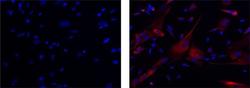
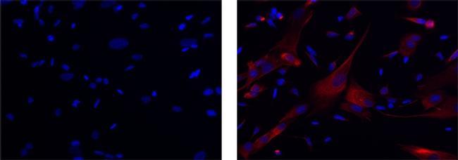
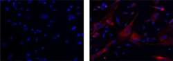
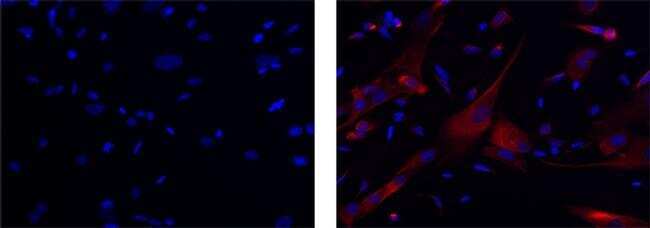
- Immunocytochemistry of MeOH-fixed SK-N-SH cells using 2.5 µg/mL of Mouse IgG1 K Isotype Control Purified (left) or 2.5 µg/mL of Anti-APP (Amyloid Precursor Protein) Purified (right) followed by F(ab')2 Anti-Mouse IgG eFluor® 570. Nuclei are stained with DAPI.
- Submitted by
- Invitrogen Antibodies (provider)
- Main image
- Experimental details
- Immunocytochemistry of MeOH-fixed SK-N-SH cells using 2.5 µg/mL of Mouse IgG1 K Isotype Control Purified (left) or 2.5 µg/mL of Anti-APP (Amyloid Precursor Protein) Purified (right) followed by F(ab')2 Anti-Mouse IgG eFluor® 570. Nuclei are stained with DAPI.
Supportive validation
- Submitted by
- Invitrogen Antibodies (provider)
- Main image
- Experimental details
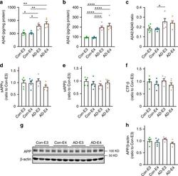
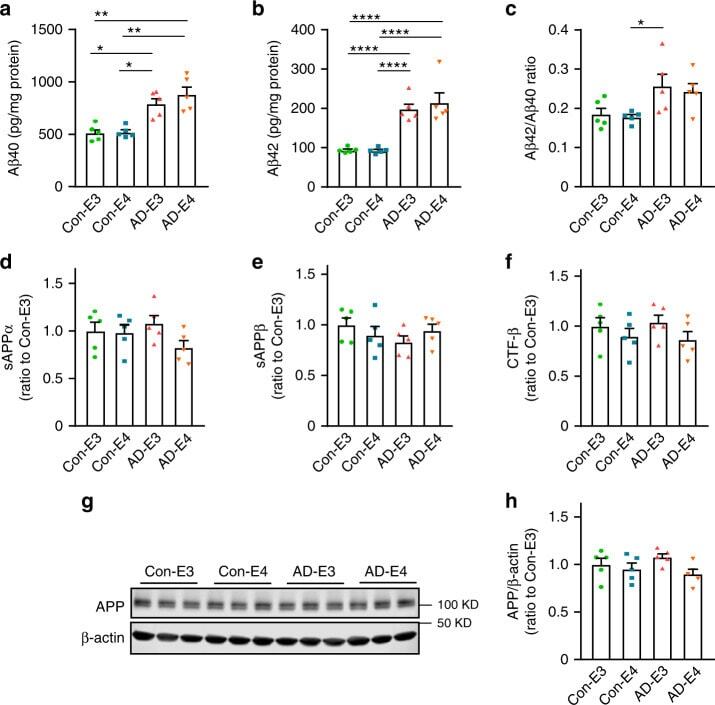
- Fig. 3 Increased Abeta accumulation in iPSC-derived cerebral organoids from AD patients. Lysates of 4-5 cerebral organoids per iPSC line were analyzed by ELISA and western blotting at week 12. a - c Amounts of Abeta40 ( a ; APOE4: p = 0.2167, AD: p = 0.0002, APOE4 x AD: p = 0.4849, Con-E3 vs. AD-E3: p = 0.0287, Con-E3 vs. AD-E4: p = 0.0028, Con-E4 vs. AD-E3: p = 0.0171, Con-E4 vs. AD-E4: p = 0.0011) and Abeta42 ( b ; APOE4: p = 0.1014, AD: p < 0.0001, APOE4 x AD: p = 0.7778, Con-E3 vs. AD-E3: p < 0.0001, Con-E3 vs. AD-E4: p < 0.0001, Con-E4 vs. AD-E3: p < 0.0001, Con-E4 vs. AD-E4: p < 0.0001) in the RIPA fraction were measured by ELISA. Data were normalized to the total protein concentration of the respective sample. The ratio of Abeta42/Abeta40 was calculated accordingly ( c ; APOE4: p = 0.9549, AD: p = 0.0034, APOE4 x AD: p = 0.5331, Con-E4 vs. AD-E3: p = 0.0397). d - f Amounts of sAPPalpha ( d ; APOE4: p = 0.1081, AD: p = 0.8009, APOE4 x AD: p = 0.1351), sAPPbeta (E; APOE4: p = 0.7772, AD: p = 0.4410, APOE4 x AD: p = 0.0936) and CTF-beta ( f ; APOE4: p = 0.1150, AD: p = 0.5326, APOE4 x AD: p = 0.9877) in RIPA were measured by ELISA. Data are shown as ratios to Con-E3 after normalization to total protein concentration. g , h Amounts of full-length APP were assessed by western blotting analysis using 22C11 monoclonal antibody in RIPA lysate ( APOE4: p = 0.0331, AD: p = 0.6706, APOE4 x AD: p = 0.3170). All data are expressed as mean +- SEM ( N = 5). ANCOVA for APOE4 , AD stat
- Submitted by
- Invitrogen Antibodies (provider)
- Main image
- Experimental details
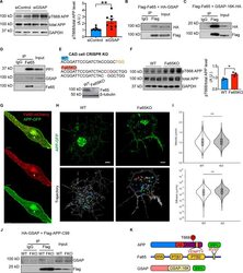
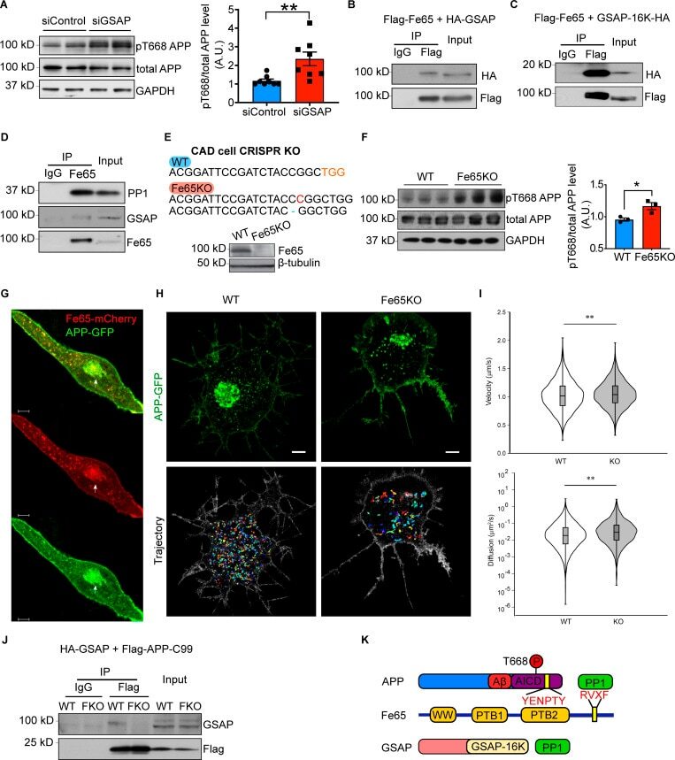
- Figure 2. GSAP interacts with Fe65 to regulate APP phosphorylation and trafficking. (A) Immunoblot analysis of protein levels in N2a695 cells transfected with control or GSAP siRNA (left panel). Quantification of APP phosphorylation at Thr668 normalized to total APP level (right panel). Data represent mean +- SEM; unpaired t test, **, P < 0.01. pT668, phospho-Thr668. Representative data of four experiments. (B) Co-IP analysis of full-length GSAP (HA-tagged) interaction with full-length Fe65 (Flag-tagged) using Flag antibody in HEK293T cells. Representative data of two experiments. (C) Co-IP analysis of GSAP C-terminal 16K domain (HA-tagged) coprecipitation with full-length Fe65 (Flag-tagged) using a Flag antibody in HEK293T cells. Representative data of two experiments. (D) Co-IP analysis of endogenous Fe65 interaction with GSAP and PP1 using Fe65 antibody in HEK293T cells. GSAP was detected using an antibody from R&D Systems. Representative data of two experiments. (E) Genomic DNA from CAD WT and Fe65KO cells was isolated, and PCR-amplified fragments flanking the CRISPR-Cas9 cleavage site were generated. PCR fragments were cloned into TOPO vector for Sanger sequencing. A 1-bp insertion (red) and deletion (blue) was identified in Fe65KO CAD cells (upper panel). Immunoblot analysis of proteins from WT and Fe65KO CAD cells (lower panel). (F) Immunoblot analysis of protein levels in CAD cells transiently overexpressing APP (left panel). Quantification of APP phosphorylation at T
- Submitted by
- Invitrogen Antibodies (provider)
- Main image

- Experimental details
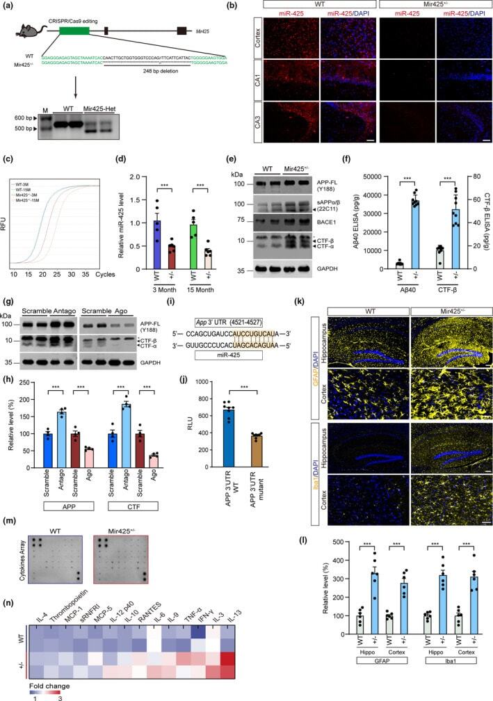
- FIGURE 3 miR-425 deficiency activates amyloidogenic APP processing and systematic neuroinflammation. (a) Generation of Mir425 +/- mice with CRISPR/Cas9 and genotypes verification of Mir425 +/- and WT mice using PCR. (b) In situ hybridization of miR-425 in hippocampus and cortex of Mir425 +/- and WT mice. Scale bars, 50 mum. (c, d) RT-PCR of miR-425 level in the hippocampus of Mir425 +/- and WT mice at 3 months and 15 month. (e) Western blot of APP, BACE1, sAPPalpha/beta, CTF-alpha, and CTF-beta in the brain of miR-425 +/- and WT mice. (f) ELISA of Abeta40 and CTF-beta in the brain of 15-month-old Mir425 +/- and WT mice. (g, h) Western blot of APP and CTF-beta in PC12 cells transfected with Antagomir-425 and Agomir-425. (i, j) Luciferase assay of miR-425 and APP 3'UTR. (k, l) Immunofluorescence and quantification of GFAP-positive and Iba1-positive glial cells in the hippocampus of 15-month-old Mir425 +/- and WT mice. Scale bars, 50 mum. (m, n) Microarray cytokines assay of hippocampal cell lysates in 15-month-old Mir425 +/- and WT mice. Data were presented as mean +- SEM. Two-tailed unpaired Student's t test. *** p < 0.001; ** p < 0.01; * p < 0.05
- Submitted by
- Invitrogen Antibodies (provider)
- Main image
- Experimental details
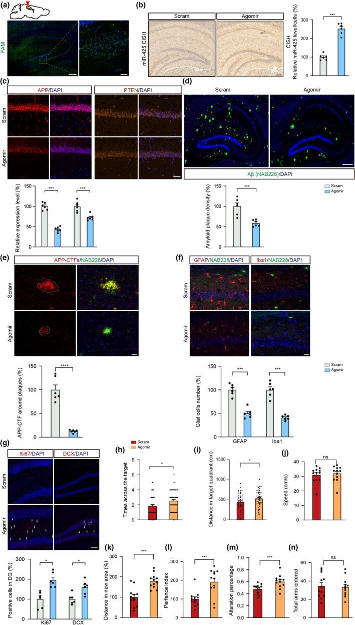
- FIGURE 7 AgomiR-425 oligonucleotide treatment ameliorates pathological and behavioral phenotypes in APP/PS1 mice. (a) Stereotactic injection of FAM-labeled oligonucleotide in the hippocampus of 6-month-old APP/PS1 mice. Scale bars, 200 mum (left), 20 mum (right). (b) Chromogenic in situ hybridization and quantification of miR-425 in hippocampus of APP/PS1 mice. Scale bars, 100 mum, n = 6 per group. (c) Immunofluorescence and quantification of APP and PTEN in AgomiR-425- and scramble-treated APP/PS1 mice, n = 6 per group. Scale bars, 50 mum. (d) Immunofluorescence and quantification of amyloid plaques in AgomiR-425- and scramble-treated APP/PS1 mice, n = 6 per group. Scale bars, 100 mum. (e, f) Immunofluorescence and quantification of APP-CTFs, GFAP and Iba1 in AgomiR-425- and scramble-treated APP/PS1 mice. Scale bars, 25 mum. (g) Immunofluorescence and quantification of Ki67- and DCX-positive neurons in AgomiR-425-and scramble-treated APP/PS1 mice. Scale bars, 50 mum. (h-j) MWM data showing times across the target, distance in target quadrant for four times and swimming speed of APP/PS1 mice, n = 12 per group. (k, l) NOR tests showing the total distance of novel object exploration and preference index of APP/PS1 mice, n = 12 per group. (m, n) Y-maze tests showing alternation percentage and the number of arm entered of APP/PS1 mice, n = 12 per group. Data were presented as mean +- SEM. Two-tailed unpaired Student's t test. *** p < 0.001; * p < 0.05