Explore
 Validate
 Learn
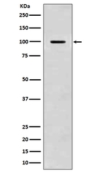
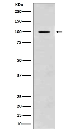
Learn Western blot
 Immunohistochemistry
Antibody data
- Antibody Data
- Antigen structure
- References [6]
- Comments [0]
- Validations
- Immunohistochemistry [1]
Submit
Validation data
Reference
Comment
Report error
- Product number
- M00081 - Provider product page

- Provider
- Boster Biological Technology
- Product name
- Anti-Amyloid beta A4 APP Rabbit Monoclonal Antibody
- Antibody type
- Monoclonal
- Description
- Monoclonal antibody for APP detection. Host: Rabbit.Size: 100ug/vial. Tested applications: Flow Cytometry, IP, IF, IHC, ICC, WB. Reactive species: Human, Mouse, Rat APP information: Molecular Weight: 86943 MW; Subcellular Localization: Membrane; Single-pass type I membrane protein. Membrane, clathrin-coated pit. Cell surface protein that rapidly becomes internalized via clathrin-coated pits. During maturation, the immature APP (N-glycosylated in the endoplasmic reticulum) moves to the Golgi complex where complete maturation occurs (O-glycosylated and sulfated). After alpha-secretase cleavage, soluble APP is released into the extracellular space and the C-terminal is internalized to endosomes and lysosomes. Some APP accumulates in secretory transport vesicles leaving the late Golgi compartment and returns to the cell surface. Gamma-CTF(59) peptide is located to both the cytoplasm and nuclei of neurons. It can be translocated to the nucleus through association with APBB1 (Fe65). Beta-APP42 associates with FRPL1 at the cell surface and the complex is then rapidly internalized. APP sorts to the basolateral surface in epithelial cells. During neuronal
- Reactivity
- Human, Mouse, Rat
- Host
- Rabbit
- Antibody clone number
- BGC-1
- Vial size
- 100ug/vial
- Concentration
- 0.5-1mg/ml, actual concentration vary by lot. Use suggested dilution ratio to decide dilution procedure.
- Storage
- At -20°C for one year. Avoid repeated freezing and thawing.
Submitted references Development of a novel bioengineered 3D brain-like tissue for studying primary blast-induced traumatic brain injury.
Severe tauopathy and axonopathy in the medulla oblongata in Alzheimer's disease implicate the changes in autonomic nervous function.
Maternal Prenatal Inflammation Increases Brain Damage Susceptibility of Lipopolysaccharide in Adult Rat Offspring via COX-2/PGD-2/DPs Pathway Activation.
α‑lipoic acid can greatly alleviate the toxic effect of AGES on SH‑SY5Y cells.
Neuroprotective Effect of Ligustilide through Induction of α-Secretase Processing of Both APP and Klotho in a Mouse Model of Alzheimer's Disease.
Lead-induced morphological changes and amyloid precursor protein accumulation in adult rat hippocampus.
Snapper DM, Reginauld B, Liaudanskaya V, Fitzpatrick V, Kim Y, Georgakoudi I, Kaplan DL, Symes AJ
Journal of neuroscience research 2023 Jan;101(1):3-19
Journal of neuroscience research 2023 Jan;101(1):3-19
Severe tauopathy and axonopathy in the medulla oblongata in Alzheimer's disease implicate the changes in autonomic nervous function.
Tian Y, Gao G, Dai J
Journal of chemical neuroanatomy 2022 Sep;123:102105
Journal of chemical neuroanatomy 2022 Sep;123:102105
Maternal Prenatal Inflammation Increases Brain Damage Susceptibility of Lipopolysaccharide in Adult Rat Offspring via COX-2/PGD-2/DPs Pathway Activation.
Zhang J, Yao P, Han W, Luo Y, Li Y, Yang Y, Xia H, Chen Z, Chen Q, Wang H, Yang L, Li H, Hu C, Huang H, Peng Z, Tan X, Li M, Yang J
International journal of molecular sciences 2022 May 30;23(11)
International journal of molecular sciences 2022 May 30;23(11)
α‑lipoic acid can greatly alleviate the toxic effect of AGES on SH‑SY5Y cells.
Niu G, Guo J, Tian Y, Zhao K, Li J, Xiao Q
International journal of molecular medicine 2018 May;41(5):2855-2864
International journal of molecular medicine 2018 May;41(5):2855-2864
Neuroprotective Effect of Ligustilide through Induction of α-Secretase Processing of Both APP and Klotho in a Mouse Model of Alzheimer's Disease.
Kuang X, Zhou HJ, Thorne AH, Chen XN, Li LJ, Du JR
Frontiers in aging neuroscience 2017;9:353
Frontiers in aging neuroscience 2017;9:353
Lead-induced morphological changes and amyloid precursor protein accumulation in adult rat hippocampus.
Sun L, Zhou XL, Yi HP, Jiang SJ, Yuan H
Biotechnic & histochemistry : official publication of the Biological Stain Commission 2014 Oct;89(7):513-7
Biotechnic & histochemistry : official publication of the Biological Stain Commission 2014 Oct;89(7):513-7
No comments: Submit comment
Supportive validation
- Submitted by
- Boster Biological Technology (provider)
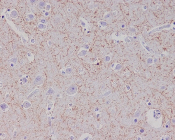
- Main image
- Experimental details
- Immunohistochemical analysis of paraffin-embedded human brain, using Amyloid beta A4 Antibody (M00081)APP was detected in paraffin-embedded tissue section. Heat mediated antigen retrieval was performed in citrate buffer (pH6, epitope retrieval solution) for 20 mins. The tissue section was blocked with 10% goat serum. The tissue section was then incubated with 1ug/ml rabbit anti-APP Antibody (M00081)overnight at 4°C. Biotinylated goat anti-rabbit IgG was used as secondary antibody and incubated for 30 minutes at 37°C. The tissue section was developed using Strepavidin-Biotin-Complex (SABC)(Catalog # SA1022) with DAB as the chromogen.
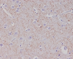
- Additional image