 Explore
 Validate
 Learn
Learn Western blot
 Immunocytochemistry
Antibody data
- Antibody Data
- Antigen structure
- References [0]
- Comments [0]
- Validations
- Immunocytochemistry [6]
- Immunoprecipitation [1]
- Other assay [1]
Submit
Validation data
Reference
Comment
Report error
- Product number
- PA5-17829 - Provider product page

- Provider
- Invitrogen Antibodies
- Product name
- Amyloid Precursor Protein Polyclonal Antibody
- Antibody type
- Polyclonal
- Antigen
- Synthetic peptide
- Description
- It is not recommended to aliquot this antibody.
- Reactivity
- Human, Mouse, Rat
- Host
- Rabbit
- Isotype
- IgG
- Vial size
- 100 μL
- Concentration
- 197 μg/mL
- Storage
- -20°C
No comments: Submit comment
Supportive validation
- Submitted by
- Invitrogen Antibodies (provider)
- Main image
- Experimental details
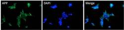
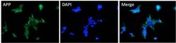
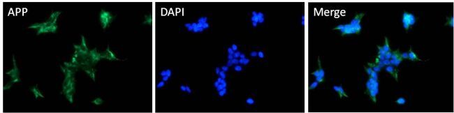
- Immunofluorescent analysis of APP (green) HEK293T cells. Cells fixed with 4% formaldehyde were permeabilized and blocked with 1X PBS containing 5% BSA and 0.3% Triton X-100 for 1 hour at room temperature. Cells were probed with an APP polyclonal antibody (Product # PA5-17829) at a dilution of 1:100 overnight at 4°C in 1X PBS containing 1% BSA and 0.3% Triton X-100, washed with 1X PBS, and incubated with a fluorophore-conjugated goat anti-rabbit IgG secondary antibody at a dilution of 1:200 for 1 hour at room temperature. Nuclei (blue) were stained with DAPI. Images were taken on a Leica DM1000 microscope at 40X magnification. Data courtesy of the Innovators Program.
- Submitted by
- Invitrogen Antibodies (provider)
- Main image

- Experimental details
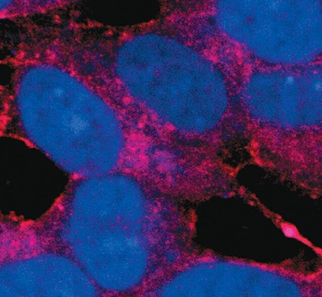
- Immunofluorescent analysis of APP in SH-SY5Y cells using an APP polyclonal antibody (Product # PA5-17829) (A) (red) and compared to an isotype control (B) showing cytoplasmic and ER staining. DNA is labeled using a fluorescent blue dye.
- Submitted by
- Invitrogen Antibodies (provider)
- Main image

- Experimental details
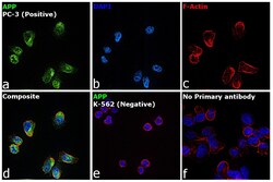
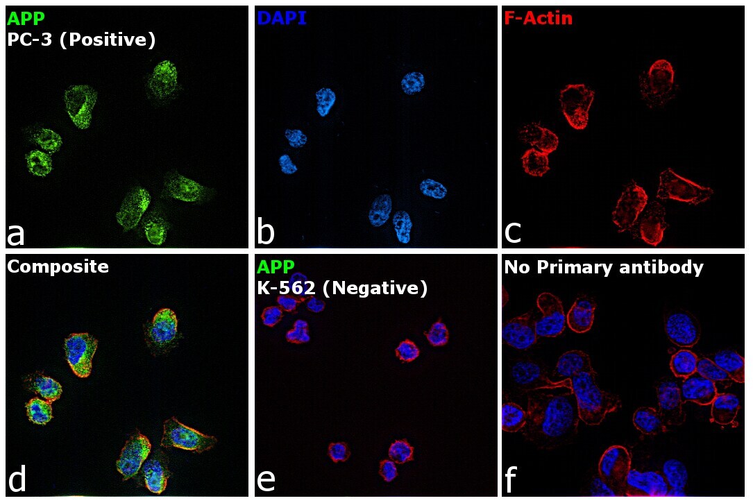
- Immunofluorescence analysis of beta Amyloid was performed using 70% confluent log phase PC-3. The cells were fixed with 4% paraformaldehyde for 10 minutes, permeabilized with 0.1% Triton™ X-100 for 15 minutes, and blocked with 2% BSA for 1 hour at room temperature. The cells were labeled with beta Amyloid Polyclonal Antibody (Product # PA5-17829) at 1:200 dilution in 0.1% BSA, incubated at 4 degree celsius overnight and then with Donkey anti-Rabbit IgG (H+L) Highly Cross-Adsorbed Secondary Antibody, Alexa Fluor Plus 488 (Product # A32790) at a dilution of 1:2000 for 45 minutes at room temperature (Panel a: green). Nuclei (Panel b: blue) were stained with ProLong™ Diamond Antifade Mountant with DAPI (Product # P36962). F-actin (Panel c: red) was stained with Rhodamine Phalloidin (Product # R415, 1:300). Panel d represents the merged image showing localization in plasma membrane, cytoplasm and nucleus. Panel e represents negative cell line K-562 with no expression. Panel f represents control cells with no primary antibody to assess background. The images were captured at 60X magnification.

- Submitted by
- Invitrogen Antibodies (provider)
- Main image

- Experimental details
- Immunofluorescent analysis of APP in SH-SY5Y cells using an APP polyclonal antibody (Product # PA5-17829) (A) (red) and compared to an isotype control (B) showing cytoplasmic and ER staining. DNA is labeled using a fluorescent blue dye.

- Submitted by
- Invitrogen Antibodies (provider)
- Main image
- Experimental details
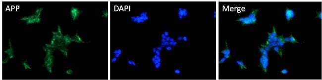
- Immunofluorescent analysis of APP (green) HEK293T cells. Cells fixed with 4% formaldehyde were permeabilized and blocked with 1X PBS containing 5% BSA and 0.3% Triton X-100 for 1 hour at room temperature. Cells were probed with an APP polyclonal antibody (Product # PA5-17829) at a dilution of 1:100 overnight at 4°C in 1X PBS containing 1% BSA and 0.3% Triton X-100, washed with 1X PBS, and incubated with a fluorophore-conjugated goat anti-rabbit IgG secondary antibody at a dilution of 1:200 for 1 hour at room temperature. Nuclei (blue) were stained with DAPI. Images were taken on a Leica DM1000 microscope at 40X magnification. Data courtesy of the Innovators Program.
- Submitted by
- Invitrogen Antibodies (provider)
- Main image
- Experimental details
- Immunofluorescence analysis of beta Amyloid was performed using 70% confluent log phase PC-3. The cells were fixed with 4% paraformaldehyde for 10 minutes, permeabilized with 0.1% Triton™ X-100 for 15 minutes, and blocked with 2% BSA for 1 hour at room temperature. The cells were labeled with beta Amyloid Polyclonal Antibody (Product # PA5-17829) at 1:200 dilution in 0.1% BSA, incubated at 4 degree celsius overnight and then with Donkey anti-Rabbit IgG (H+L) Highly Cross-Adsorbed Secondary Antibody, Alexa Fluor Plus 488 (Product # A32790) at a dilution of 1:2000 for 45 minutes at room temperature (Panel a: green). Nuclei (Panel b: blue) were stained with ProLong™ Diamond Antifade Mountant with DAPI (Product # P36962). F-actin (Panel c: red) was stained with Rhodamine Phalloidin (Product # R415, 1:300). Panel d represents the merged image showing localization in plasma membrane, cytoplasm and nucleus. Panel e represents negative cell line K-562 with no expression. Panel f represents control cells with no primary antibody to assess background. The images were captured at 60X magnification.
Supportive validation
- Submitted by
- Invitrogen Antibodies (provider)
- Main image

- Experimental details
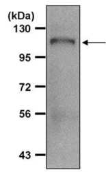
- Immunoprecipitation of APP was performed on HEK293T cells. Antigen-antibody complexes were formed by incubating 500 µg of HEK293T whole cell lysate (in 500 µL volume) with 5 µL of an APP polyclonal antibody (Product # PA5-17829) overnight at 4°C. The immune complexes were captured on 30 µL of protein G agarose, washed extensively, and eluted with 6X Laemmli buffer. Samples were resolved on an 8% SDS-PAGE gel, transferred to a PVDF membrane, and blocked with 5% milk in TBST for 1 hour at room temperature. The membrane was probed with an APP polyclonal antibody (Product # PA5-17829) at a dilution of 1:1000 overnight at 4°C, washed in TBST, and probed with an HRP-conjugated mouse anti-rabbit light chain specific secondary antibody at a dilution of 1:40,000 for 1 hour at room temperature. Chemiluminescent detection was performed using ECL substrate. Data courtesy of the Innovators Program.

Supportive validation
- Submitted by
- Invitrogen Antibodies (provider)
- Main image
- Experimental details
- Immunoprecipitation of APP was performed on HEK293T cells. Antigen-antibody complexes were formed by incubating 500 µg of HEK293T whole cell lysate (in 500 µL volume) with 5 µL of an APP polyclonal antibody (Product # PA5-17829) overnight at 4°C. The immune complexes were captured on 30 µL of protein G agarose, washed extensively, and eluted with 6X Laemmli buffer. Samples were resolved on an 8% SDS-PAGE gel, transferred to a PVDF membrane, and blocked with 5% milk in TBST for 1 hour at room temperature. The membrane was probed with an APP polyclonal antibody (Product # PA5-17829) at a dilution of 1:1000 overnight at 4°C, washed in TBST, and probed with an HRP-conjugated mouse anti-rabbit light chain specific secondary antibody at a dilution of 1:40,000 for 1 hour at room temperature. Chemiluminescent detection was performed using ECL substrate. Data courtesy of the Innovators Program.
