Explore
 Validate
 Learn
Learn Western blot
 ELISA
ELISAAntibody data
- Antibody Data
- Antigen structure
- References [0]
- Comments [0]
- Validations
- Western blot [3]
- Immunohistochemistry [2]
Submit
Validation data
Reference
Comment
Report error
- Product number
- GTX82974 - Provider product page

- Provider
- GeneTex
- Proper citation
- GeneTex Cat#GTX82974, RRID:AB_1240447
- Product name
- beta Amyloid antibody
- Antibody type
- Polyclonal
- Reactivity
- Human, Mouse
- Host
- Rabbit
No comments: Submit comment
Supportive validation
- Submitted by
- GeneTex (provider)
- Main image
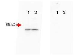
- Experimental details
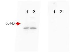
- Mouse Brain (Lane 1) and Mouse Spinal Chord (Lane 2) were run on a 4-20% gradient gel, Blocked in 1% BSA-TBS-T 30 min RT and probed with Rb-a-Beta Amyloid (GTX82974) 1:1000 in 1% BSA-TBS-T o/n 4°C. HRP Gt-a-Rb 611-103-122 Lot#21231 1:40,000 in blocking buffer 30 min RT. FEMTOMAX chemiluminescent substrate was used for detection of a 40-50 kD band consistent with a higher MW precursor form of beta amyloid. A secondary Ab only control (Shown right) showed no detectable signal.
- Validation comment
- WB
- Submitted by
- GeneTex (provider)
- Main image
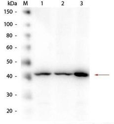
- Experimental details
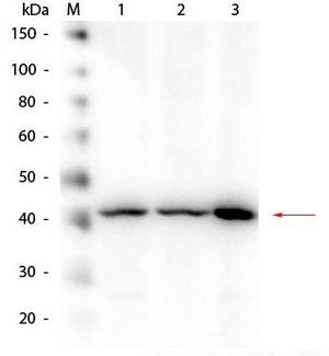
- Western Blot of Rabbit anti-Beta Amyloid Antibody. Lane 1: HEK293 WCL. Lane 2: Mouse Brain WCL. Lane 3: A-172 WCL. Load: 10.0 ?g per lane. Primary antibody: Beta Amyloid Antibody at 1:1,000 overnight at 4¢XC. Secondary antibody: Peroxidase Conjugated Goat-a-Rabbit IgG at 1:40,000 for 30 min at RT. Block for 30 min at RT. Predicted/Observed size: 40 kDa, 40 kDa for Beta Amyloid.
- Submitted by
- GeneTex (provider)
- Main image
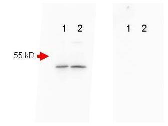
- Experimental details
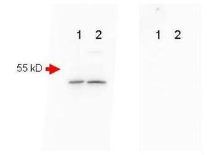
- Mouse Brain (Lane 1) and Mouse Spinal Chord (Lane 2) were run on a 4-20% gradient gel, Blocked in 1% BSA-TBS-T 30 min RT and probed with Rb-a-Beta Amyloid (GTX82974) 1:1000 in 1% BSA-TBS-T o/n 4¢XC. FEMTOMAX chemiluminescent substrate was used for detection of a 40-50 kD band consistent with a higher MW precursor form of beta amyloid. A secondary Ab only control (Shown right) showed no detectable signal.
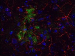
Supportive validation
- Submitted by
- GeneTex (provider)
- Main image
- Experimental details
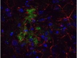
- Immunohistochemistical detection of beta Amyloid using Anti-Beta Amyloid Antibody on TG APP23 mouse brain cortex frozen sections. Anti-Beta Amyloid Antibody used at 1/200 and incubated for 2 hours in TBS/BSA/Tween/azide. Fluorescent labelled anti rabbit IgG was then added.
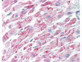
- Submitted by
- GeneTex (provider)
- Main image
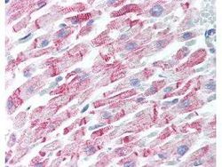
- Experimental details
- Human Heart (formalin-fixed, paraffin-embedded) stained with Anti-Beta Amyloid Antibody (GTX82974) at 5 ug/ml followed by biotinylated goat anti-rabbit IgG secondary antibody, alkaline phosphatase-streptavidin and chromogen.