 Explore
 Validate
 Learn
Learn Western blot
Antibody data
- Antibody Data
- Antigen structure
- References [0]
- Comments [0]
- Validations
- Western blot [1]
- Immunocytochemistry [1]
- Immunohistochemistry [1]
Submit
Validation data
Reference
Comment
Report error
- Product number
- MAB2432-100 - Provider product page

- Provider
- R&D Systems
- Product name
- Human TAP2 Antibody
- Antibody type
- Monoclonal
- Description
- Protein A or G purified from ascites. Detects human TAP2 in direct ELISAs.
- Reactivity
- Human
- Host
- Mouse
- Conjugate
- Unconjugated
- Antigen sequence
Q03519- Isotype
- IgG
- Antibody clone number
- 998524
- Vial size
- 100 ug
- Storage
- Use a manual defrost freezer and avoid repeated freeze-thaw cycles. 12 months from date of receipt, -20 to -70 °C as supplied. 1 month, 2 to 8 °C under sterile conditions after reconstitution. 6 months, -20 to -70 °C under sterile conditions after reconstitution.
No comments: Submit comment
Supportive validation
- Submitted by
- R&D Systems (provider)
- Main image
- Experimental details
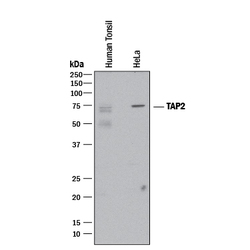
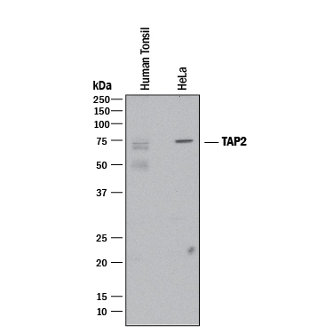
- Detection of Human TAP2 by Western Blot. Western blot shows lysates of human tonsil tissue and HeLa human cervical epithelial carcinoma cell line. PVDF membrane was probed with 2 µg/mL of Mouse Anti-Human TAP2 Monoclonal Antibody (Catalog # MAB2432) followed by HRP-conjugated Anti-Mouse IgG Secondary Antibody (Catalog # HAF018). A specific band was detected for TAP2 at approximately 75 kDa (as indicated). This experiment was conducted under reducing conditions and using Immunoblot Buffer Group 1.
Supportive validation
- Submitted by
- R&D Systems (provider)
- Main image

- Experimental details
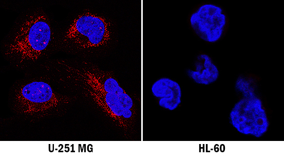
- TAP2 in U-251 MG and HL-60 Human Cell Line. TAP2 was detected in immersion fixed U-251 MG human glioblastoma cell line (left panel) and HL-60 human acute promyelocytic leukemia cell line (right panel, negative control) using Mouse Anti-Human TAP2 Monoclonal Antibody (Catalog # MAB2432) at 8 µg/mL for 3 hours at room temperature. Cells were stained using the NorthernLights™ 557-conjugated Anti-Mouse IgG Secondary Antibody (red; Catalog # NL007) and counterstained with DAPI (blue). Specific staining was localized to cytoplasm. View our protocol for Fluorescent ICC Staining of Cells on Coverslips.
Supportive validation
- Submitted by
- R&D Systems (provider)
- Main image
- Experimental details
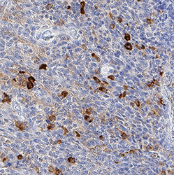
- TAP2 in Human Tonsil. TAP2 was detected in immersion fixed paraffin-embedded sections of human tonsil using Mouse Anti-Human TAP2 Monoclonal Antibody (Catalog # MAB2432) at 8 µg/mL for 1 hour at room temperature followed by incubation with the Anti-Mouse IgG VisUCyte™ HRP Polymer Antibody (Catalog # VC001). Before incubation with the primary antibody, tissue was subjected to heat-induced epitope retrieval using Antigen Retrieval Reagent-Basic (Catalog # CTS013). Tissue was stained using DAB (brown) and counterstained with hematoxylin (blue). Specific staining was localized to cytoplasm in lymphocytes. View our protocol for IHC Staining with VisUCyte HRP Polymer Detection Reagents.
