 Explore
 Validate
 Learn
Learn Western blot
 Immunocytochemistry
Antibody data
- Antibody Data
- Antigen structure
- References [0]
- Comments [0]
- Validations
- Western blot [4]
- Immunohistochemistry [1]
- Flow cytometry [1]
Submit
Validation data
Reference
Comment
Report error
- Product number
- NBP2-47774-0.1mg - Provider product page

- Provider
- Novus Biologicals
- Product name
- Mouse Monoclonal ZAP70 Antibody
- Antibody type
- Monoclonal
- Description
- Protein A or G purified. ZAP70 is a 70kDa protein tyrosine kinase found in T-cells and natural killer cells. Control of this protein translation is via the IgVH gene. In Western blotting of whole cell lysates of normal peripheral blood mononuclear cells, the antibody labels a band corresponding to ZAP70. In Western blotting of whole cell lysates of CD19-positive purified leukemia cells from patients with Ig-unmutated and Ig-mutated CLL, the antibody labels a band corresponding to ZAP70 in the Ig-unmutated CLL samples, whereas no band is observed in the Ig-mutated CLL samples. In Western blotting of cell lysates of Jurkat cells (T-lymphoblastic cell line), the antibody labels a band of 70kDa protein. In Western blotting of cell lysates of A431 cells (carcinoma cell line), no band is observed. ZAP70 protein is expressed in leukemic cells of approximately 25% of chronic lymphocytic leukemia (CLL) cases as well. Anti-ZAP70 expression is an excellent surrogate marker for the distinction between the Ig-mutated (anti-ZAP70 negative) and Ig-unmutated (anti-ZAP70 positive) CLL subtypes and can identify patient groups with divergent clinical courses. The anti-ZAP70 positive Ig-unmutated CLL cases have been shown to have a poorer prognosis.
- Reactivity
- Human
- Host
- Mouse
- Isotype
- IgG
- Vial size
- 0.1 mg
- Concentration
- 1.0 mg/ml
- Storage
- Store at 4C short term. Aliquot and store at -20C long term. Avoid freeze-thaw cycles.
No comments: Submit comment
Supportive validation
- Submitted by
- Novus Biologicals (provider)
- Main image

- Experimental details
- Simple Western: ZAP70 Antibody (2F3.2) - Azide and BSA Free [NBP2-47774] - Lane view shows a specific band for ZAP70 in Jurkat cell lysate using 50ug/ml antibody dilution. Electropherogram image of corresponding Simple Western lane view at WES molecular weight of 70. Image from an internal validation.

- Submitted by
- Novus Biologicals (provider)
- Main image

- Experimental details
- Simple Western: ZAP70 Antibody (2F3.2) - Azide and BSA Free [NBP2-47774] - Electropherogram image of the corresponding Simple Western lane. ZAP70 antibody was used at 10 ug/ml dilution of Jurkat and H. Tonsil lysates(s) respectively.

- Submitted by
- Novus Biologicals (provider)
- Main image

- Experimental details
- Simple Western: ZAP70 Antibody (2F3.2) - Azide and BSA Free [NBP2-47774] - Simple Western lane view shows a specific band for ZAP70 in 0.2 mg/ml of Jurkat (left) and H. Tonsil (right) lysate(s). This experiment was performed under reducing conditions using the 12-230 kDa separation system.

- Submitted by
- Novus Biologicals (provider)
- Main image

- Experimental details
- Simple Western: ZAP70 Antibody (2F3.2) - Azide and BSA Free [NBP2-47774] - Lane view shows a specific band for ZAP70 in Jurkat cell lysate using 50 ug/mL antibody dilution. Electropherogram image of corresponding Simple Western lane view at WES molecular weight of 70 kDa. Image from an internal validation.

Supportive validation
- Submitted by
- Novus Biologicals (provider)
- Main image

- Experimental details
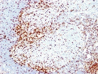
- Immunohistochemistry-Paraffin: ZAP70 Antibody (2F3.2) - Azide and BSA Free [NBP2-47774] - Human Tonsil stained with ZAP70 Monoclonal Antibody (2F3.2).
Supportive validation
- Submitted by
- Novus Biologicals (provider)
- Main image

- Experimental details
- Flow Cytometry: ZAP70 Antibody (2F3.2) - Azide and BSA Free [NBP2-47774] - Flow Cytometric Analysis of PFA-fixed Jurkat cells. ZAP70 Antibody (2F3.2) followed by goat anti-Mouse IgG-CF488 (Blue); Isotype Control (Red).
