Explore
 Validate
 Learn
Learn Western blot
 Immunoprecipitation
Antibody data
- Antibody Data
- Antigen structure
- References [5]
- Comments [0]
- Validations
- Western blot [2]
- Immunocytochemistry [1]
- Flow cytometry [1]
Submit
Validation data
Reference
Comment
Report error
- Product number
- 14-6695-82 - Provider product page

- Provider
- Invitrogen Antibodies
- Product name
- Zap-70 Monoclonal Antibody (1E7.2), eBioscience™
- Antibody type
- Monoclonal
- Antigen
- Other
- Description
- Description: The 1E7.2 antibody reacts with human and mouse ZAP-70, the TCRζ-associated protein-70. ZAP-70 is a cytosolic protein tyrosine kinase (PTK) and a member of the Syk family of proteins. It is expressed in T and NK cells and is required for TCR signaling and development. ZAP-70 interacts with the TCR complex by binding to tyrosine-phosphorylated immunoreceptor tyrosine-based activation motifs (ITAMs) present in the invariant subunits of the TCR complex. Following activation, ZAP-70 is phosphorylated on several tyrosine residues by two mechanisms; an autophosphorylation and a transphosphorylation by the Src family tyrosine kinase Lck1-3. Tyrosine phosphorylation of ZAP-70 correlates to its increased kinase activity and triggers downstream signaling events. Mutations in ZAP-70 have been shown to result in a form of Severe Combined Immunodeficiency Syndrome (SCID) in humans. 1E7.2 was generated against a KLH-peptide sequence corresponding to the human ZAP-70 amino acid residues 282-307. While ZAP-70 is normally expressed in T and NK cells, several recent studies have also shown high correlation of ZAP-70 positive expression with mutated IgVH expression in B-chronic lymphocytic leukemia (CCL). In conclusion, the expression of ZAP-70, which can be measured by intracellular flow cytometry, may serve as a prognostic marker for B-CLL. Applications Reported: The 1E7.2 antibody has been reported for use in intracellular staining followed by flow cytometric analysis, immunoprecipitation, and immunoblotting (WB). (Fluorochrome conjugated 1E7.2 is recommended for use in intracellular staining and flow cytometric analysis.). Applications Tested: This 1E7.2 antibody has been tested by immunoblotting of mouse thymocyte and human Jurkat cell lysates. This can be used at less than or equal to 1 µg per test. A test is defined as the amount (µg) of antibody that will stain a cell sample in a final volume of 100 µL. Cell number should be determined empirically but can range from 10^5 to 10^8 cells/test. It is recommended that the antibody be carefully titrated for optimal performance in the assay of interest. Purity: Greater than 90%, as determined by SDS-PAGE. Aggregation: Less than 10%, as determined by HPLC. Filtration: 0.2 µm post-manufacturing filtered.
- Reactivity
- Human, Mouse
- Host
- Mouse
- Isotype
- IgG
- Antibody clone number
- 1E7.2
- Vial size
- 100 µg
- Concentration
- 0.5 mg/mL
- Storage
- 4° C
Submitted references Ectopically expressed PIR-B on T cells constitutively binds to MHC class I and attenuates T helper type 1 responses.
ZAP-70 expression and prognosis in chronic lymphocytic leukaemia.
Expression of ZAP-70 is associated with increased B-cell receptor signaling in chronic lymphocytic leukemia.
Tyrosine phosphorylation of Pyk2 is selectively regulated by Fyn during TCR signaling.
Dominant-negative zeta-associated protein 70 inhibits T cell antigen receptor signaling.
Imada M, Masuda K, Satoh R, Ito Y, Goto Y, Matsuoka T, Endo S, Nakamura A, Kawamoto H, Takai T
International immunology 2009 Oct;21(10):1151-61
International immunology 2009 Oct;21(10):1151-61
ZAP-70 expression and prognosis in chronic lymphocytic leukaemia.
Orchard JA, Ibbotson RE, Davis Z, Wiestner A, Rosenwald A, Thomas PW, Hamblin TJ, Staudt LM, Oscier DG
Lancet (London, England) 2004 Jan 10;363(9403):105-11
Lancet (London, England) 2004 Jan 10;363(9403):105-11
Expression of ZAP-70 is associated with increased B-cell receptor signaling in chronic lymphocytic leukemia.
Chen L, Widhopf G, Huynh L, Rassenti L, Rai KR, Weiss A, Kipps TJ
Blood 2002 Dec 15;100(13):4609-14
Blood 2002 Dec 15;100(13):4609-14
Tyrosine phosphorylation of Pyk2 is selectively regulated by Fyn during TCR signaling.
Qian D, Lev S, van Oers NS, Dikic I, Schlessinger J, Weiss A
The Journal of experimental medicine 1997 Apr 7;185(7):1253-9
The Journal of experimental medicine 1997 Apr 7;185(7):1253-9
Dominant-negative zeta-associated protein 70 inhibits T cell antigen receptor signaling.
Qian D, Mollenauer MN, Weiss A
The Journal of experimental medicine 1996 Feb 1;183(2):611-20
The Journal of experimental medicine 1996 Feb 1;183(2):611-20
No comments: Submit comment
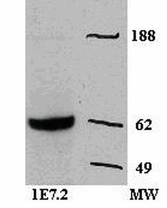
Supportive validation
- Submitted by
- Invitrogen Antibodies (provider)
- Main image
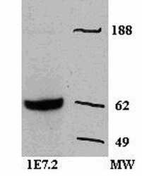
- Experimental details
- Immunoblotting of Jurkat cell lysate from 5x10e7 cells/mL (probed with 2 µg/mL of Anti-Human/Mouse ZAP-70 Purified).
- Submitted by
- Invitrogen Antibodies (provider)
- Main image

- Experimental details
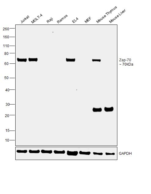
- Western blot was performed using Anti-Zap-70 Monoclonal Antibody (1E7.2), eBioscience™(Product # 14-6695-82) and a 70kDa band corresponding to Zap-70 was observed across cell lines and tissue tested. Membrane enriched extracts (30 µg lysate) of Jurkat (Lane 1), MOLT-4 (Lane 2), Raji (Lane 3), Ramos (Lane 4), EL4 (Lane 5) and MEF (Lane 6). Tissue Extracts of Mouse Thymus (Lane 7) and Mouse Liver (Lane 8) were electrophoresed using NuPAGE™ 4-12% Bis-Tris Protein Gel (Product # NP0321BOX). Resolved proteins were then transferred onto a Nitrocellulose membrane (Product # IB23002) by iBlot® 2 Dry Blotting System (Product # IB21001). The blot was probed with the primary antibody (2ug/ml) and detected by chemiluminescence with Goat anti-Mouse IgG (H+L) Superclonal™ Recombinant Secondary Antibody, HRP (Product # A28177, 1:4000 dilution) using the iBright FL 1000 (Product # A32752). Chemiluminescent detection was performed using Novex® ECL Chemiluminescent Substrate Reagent Kit (Product # WP20005).
Supportive validation
- Submitted by
- Invitrogen Antibodies (provider)
- Main image
- Experimental details
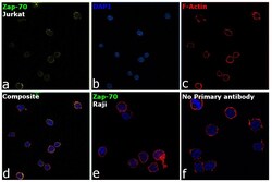
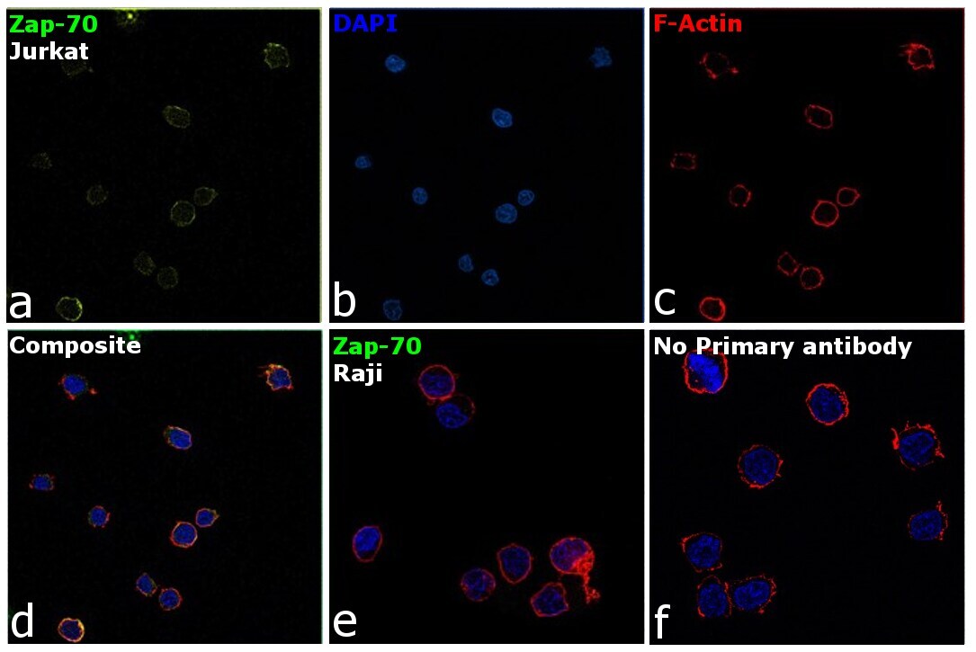
- Immunofluorescence analysis of Zap-70 was performed using 70 confluent log phase Jurkat cells. The cells were fixed with 4% paraformaldehyde for 10 minutes, permeabilized with 0.1% Triton™ X-100 for 15 minutes, and blocked with 2% BSA for 45 minutes at room temperature. The cells were labeled with Zap-70 Monoclonal Antibody (1E7.2), eBioscience™ (Product # 14-6695-82) at 5 µg/mL in 0.1% BSA, incubated at 4 degree celsius overnight and then labeled with Donkey anti-Mouse IgG (H+L) Highly Cross-Adsorbed Secondary Antibody, Alexa Fluor Plus 647 (Product # A32787), (1:2000 dilution), for 45 minutes at room temperature (Panel a: Green). Nuclei (Panel b: Blue) were stained with ProLong™ Diamond Antifade Mountant with DAPI (Product # P36962). F-actin (Panel c: Red) was stained with Rhodamine Phalloidin (Product # R415, 1:300 dilution). Panel d represents the merged image showing membrane localization. Panel e represents Raji cells with no expression of Zap-70. Panel f represents control cells with no primary antibody to assess background. The images were captured at 60X magnification.
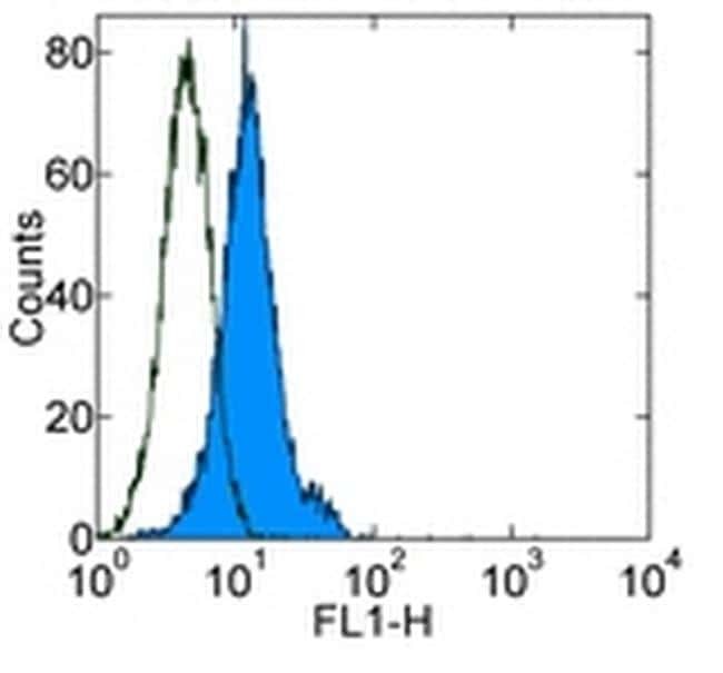
Supportive validation
- Submitted by
- Invitrogen Antibodies (provider)
- Main image

- Experimental details
- Staining of permeabilized Jurkat cells with 0.5 µg of Mouse IgG1 K Isotype Control Purified (Product # 14-4714-82) (open histogram) or 0.5 µg of Anti-Human/Mouse ZAP-70 Purified (filled histogram) followed by Anti-Mouse IgG FITC (Product # 11-4011-85).Total cells were used for analysis.