 Explore
 Validate
 Learn
Learn Flow cytometry
Antibody data
- Antibody Data
- Antigen structure
- References [1]
- Comments [0]
- Validations
- Flow cytometry [1]
Submit
Validation data
Reference
Comment
Report error
- Product number
- 51-6695-82 - Provider product page

- Provider
- Invitrogen Antibodies
- Product name
- ZAP-70 Monoclonal Antibody (1E7.2), Alexa Fluor™ 647, eBioscience™
- Antibody type
- Monoclonal
- Antigen
- Other
- Description
- Description: The 1E7.2 antibody reacts with human and mouse ZAP-70, the TCR zeta-associated protein-70. ZAP-70 is a cytosolic protein tyrosine kinase (PTK) and a member of the Syk family of proteins. It is expressed in T and NK cells and is required for TCR signaling and development. ZAP-70 interacts with the TCR complex by binding to tyrosine-phosphorylated immunoreceptor tyrosine-based activation motifs (ITAMs) present in the invariant subunits of the TCR complex. Following activation, ZAP-70 is phosphorylated on several tyrosine residues by two mechanisms; an autophosphorylation and a transphosphorylation by the Src family tyrosine kinase Lck1-3. Tyrosine phosphorylation of ZAP-70 correlates to its increased kinase activity and triggers downstream signaling events. Mutations in ZAP-70 have been shown to result in a form of Severe Combined Immunodeficiency Syndrome (SCID) in humans. 1E7.2 was generated against a KLH-peptide sequence corresponding to the human ZAP-70 amino acid residues 282-307. While ZAP-70 is normally expressed in T and NK cells, several recent studies have also shown high correlation of ZAP-70 positive expression with mutated IgVH expression in B-chronic lymphocytic leukemia (CCL). In conclusion, the expression of ZAP-70, which can be measured by intracellular flow cytometry, may serve as a prognostic marker for B-CLL. Applications Reported: The 1E7.2 antibody has been reported for use in intracellular flow cytometric analysis. Applications Tested: This 1E7.2 antibody has been tested by intracellular flow cytometric analysis on normal human peripheral blood cells using the Foxp3/Transcription Factor Buffer Set (Product # 00-5523-00) and protocol. Please refer to BestProtocols®: Protocol B: One step protocol for (nuclear) intracellular proteins. This can be used at less than or equal to 0.5 µg per test. A test is defined as the amount (µg) of antibody that will stain a cell sample in a final volume of 100 µL. Cell number should be determined empirically but can range from 10^5 to 10^8 cells/test. It is recommended that the antibody be carefully titrated for optimal performance in the assay of interest. Excitation: 633-647 nm; Emission: 668 nm; Laser: Red Laser.
- Reactivity
- Human, Mouse
- Host
- Mouse
- Conjugate
- Red dye
- Isotype
- IgG
- Antibody clone number
- 1E7.2
- Vial size
- 100 μg
- Concentration
- 0.2 mg/mL
- Storage
- 4°C, store in dark, DO NOT FREEZE!
Submitted references Temporal analysis of T-cell receptor-imposed forces via quantitative single molecule FRET measurements.
Göhring J, Kellner F, Schrangl L, Platzer R, Klotzsch E, Stockinger H, Huppa JB, Schütz GJ
Nature communications 2021 May 4;12(1):2502
Nature communications 2021 May 4;12(1):2502
No comments: Submit comment
Supportive validation
- Submitted by
- Invitrogen Antibodies (provider)
- Main image
- Experimental details
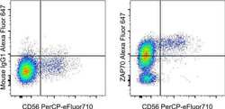
- Normal human peripheral blood cells were stained with CD56 Monoclonal Antibody, PerCP-eFluor710 (Product # 46-0566-42) and Mouse IgG1 kappa Isotype Control, Alexa Fluor 647 (Product # 51-4714-81) (left) or ZAP70 Monoclonal Antibody, Alexa Fluor 647 (right). Cells in the lymphocyte gate were used for analysis.
