 Explore
 Validate
 Learn
Learn Western blot
 Immunocytochemistry
Antibody data
- Antibody Data
- Antigen structure
- References [0]
- Comments [0]
- Validations
- Immunocytochemistry [2]
- Flow cytometry [6]
Submit
Validation data
Reference
Comment
Report error
- Product number
- MA1-19443 - Provider product page

- Provider
- Invitrogen Antibodies
- Product name
- Zap-70 Monoclonal Antibody (ZAP-03)
- Antibody type
- Monoclonal
- Antigen
- Other
- Description
- This antibody reacts with ZAP70, a 70 kDa protein tyrosine kinase expressed in T and NK cells (intracellular antigen). ZAP70 is a molecule susceptible to degradation. It is recommended to use freshly prepared cell lysates (protease inhibitors are essential) to avoid non-specific staining of degradation products.
- Reactivity
- Human, Mouse
- Host
- Mouse
- Isotype
- IgG
- Antibody clone number
- ZAP-03
- Vial size
- 100 μg
- Concentration
- 1 mg/mL
- Storage
- 4°C, do not freeze
No comments: Submit comment
Supportive validation
- Submitted by
- Invitrogen Antibodies (provider)
- Main image

- Experimental details
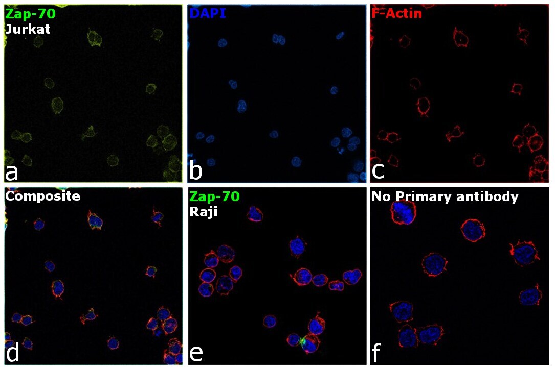
- Immunofluorescence analysis of Zap-70 was performed using 70 confluent log phase Jurkat cells. The cells were fixed with 4% paraformaldehyde for 10 minutes, permeabilized with 0.1% Triton™ X-100 for 15 minutes, and blocked with 2% BSA for 45 minutes at room temperature. The cells were labeled with Zap-70 Monoclonal Antibody (ZAP-03) (Product # MA1-19443) at 1:100 dilution in 0.1% BSA, incubated at 4 degree celsius overnight and then labeled with Donkey anti-Mouse IgG (H+L) Highly Cross-Adsorbed Secondary Antibody, Alexa Fluor Plus 647 (Product # A32787), (1:2000 dilution), for 45 minutes at room temperature (Panel a: Green). Nuclei (Panel b: Blue) were stained with ProLong™ Diamond Antifade Mountant with DAPI (Product # P36962). F-actin (Panel c: Red) was stained with Rhodamine Phalloidin (Product # R415, 1:300 dilution). Panel d represents the merged image showing membrane localization. Panel e represents Raji cells with no expression of Zap-70. Panel f represents control cells with no primary antibody to assess background. The images were captured at 60X magnification.
- Submitted by
- Invitrogen Antibodies (provider)
- Main image
- Experimental details
- Immunofluorescence analysis of Zap-70 was performed using 70 confluent log phase Jurkat cells. The cells were fixed with 4% paraformaldehyde for 10 minutes, permeabilized with 0.1% Triton™ X-100 for 15 minutes, and blocked with 2% BSA for 45 minutes at room temperature. The cells were labeled with Zap-70 Monoclonal Antibody (ZAP-03) (Product # MA1-19443) at 1:100 dilution in 0.1% BSA, incubated at 4 degree celsius overnight and then labeled with Donkey anti-Mouse IgG (H+L) Highly Cross-Adsorbed Secondary Antibody, Alexa Fluor Plus 647 (Product # A32787), (1:2000 dilution), for 45 minutes at room temperature (Panel a: Green). Nuclei (Panel b: Blue) were stained with ProLong™ Diamond Antifade Mountant with DAPI (Product # P36962). F-actin (Panel c: Red) was stained with Rhodamine Phalloidin (Product # R415, 1:300 dilution). Panel d represents the merged image showing membrane localization. Panel e represents Raji cells with no expression of Zap-70. Panel f represents control cells with no primary antibody to assess background. The images were captured at 60X magnification.

Supportive validation
- Submitted by
- Invitrogen Antibodies (provider)
- Main image

- Experimental details
- Flow cytometry intracellular staining pattern of human peripheral whole blood using anti-ZAP70 (ZAP-03) purified Monoclonal antibody (Product # MA1-19443) (concentration in sample 9 µg/mL, GAM APC).

- Submitted by
- Invitrogen Antibodies (provider)
- Main image

- Experimental details
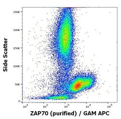
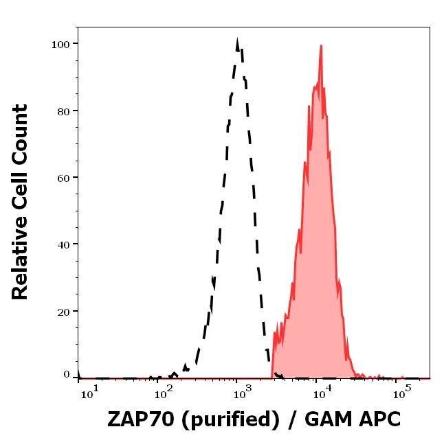
- Separation of human CD3 negative ZAP70 positive lymphocytes (red-filled) from neutrophil granulocytes (black-dashed) in flow cytometry analysis (intracellular staining) of peripheral whole blood stained using anti-ZAP70 (ZAP-03) purified Monoclonal antibody (Product # MA1-19443) (concentration in sample 9 µg/mL, GAM APC).

- Submitted by
- Invitrogen Antibodies (provider)
- Main image

- Experimental details
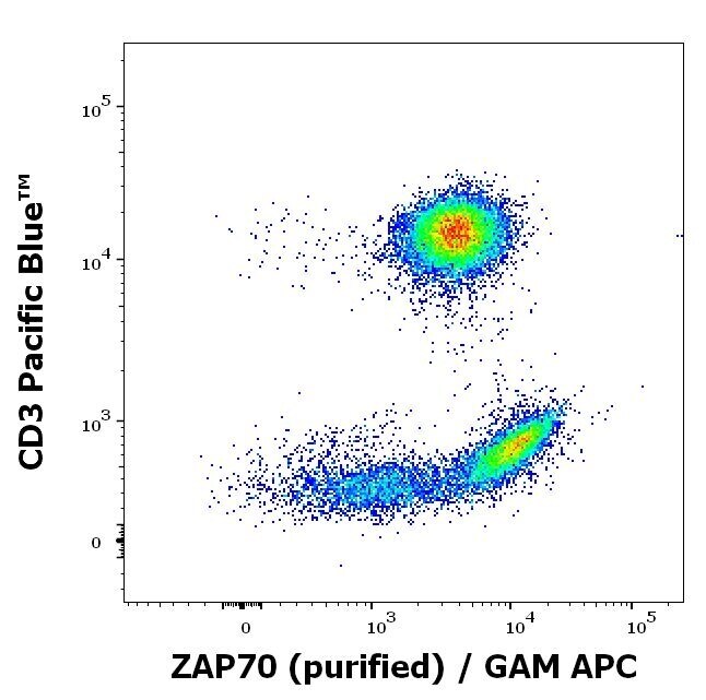
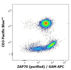
- Flow cytometry multicolor intracellular staining of human peripheral whole blood stained using anti-ZAP70 (ZAP-03) purified Monoclonal antibody (Product # MA1-19443) (concentration in sample 9 µg/mL, GAM APC) and anti-human CD3 (UCHT1) Pacific Blue™ using a dilution of 20 µL reagent/100 µL of peripheral whole blood.
- Submitted by
- Invitrogen Antibodies (provider)
- Main image
- Experimental details
- Flow cytometry intracellular staining pattern of human peripheral whole blood using anti-ZAP70 (ZAP-03) purified Monoclonal antibody (Product # MA1-19443) (concentration in sample 9 µg/mL, GAM APC).

- Submitted by
- Invitrogen Antibodies (provider)
- Main image

- Experimental details
- Separation of human CD3 negative ZAP70 positive lymphocytes (red-filled) from neutrophil granulocytes (black-dashed) in flow cytometry analysis (intracellular staining) of peripheral whole blood stained using anti-ZAP70 (ZAP-03) purified Monoclonal antibody (Product # MA1-19443) (concentration in sample 9 µg/mL, GAM APC).
- Submitted by
- Invitrogen Antibodies (provider)
- Main image
- Experimental details
- Flow cytometry multicolor intracellular staining of human peripheral whole blood stained using anti-ZAP70 (ZAP-03) purified Monoclonal antibody (Product # MA1-19443) (concentration in sample 9 µg/mL, GAM APC) and anti-human CD3 (UCHT1) Pacific Blue™ using a dilution of 20 µL reagent/100 µL of peripheral whole blood.
