Explore
 Validate
 Learn
Learn Western blot
Antibody data
- Antibody Data
- Antigen structure
- References [7]
- Comments [0]
- Validations
- Western blot [2]
- Immunocytochemistry [1]
- Immunohistochemistry [2]
- Other assay [4]
Submit
Validation data
Reference
Comment
Report error
- Product number
- 700349 - Provider product page

- Provider
- Invitrogen Antibodies
- Product name
- Phospho-STAT1 (Tyr701) Recombinant Rabbit Monoclonal Antibody (15H13L67)
- Antibody type
- Monoclonal
- Antigen
- Synthetic peptide
- Reactivity
- Human, Mouse
- Host
- Rabbit
- Isotype
- IgG
- Antibody clone number
- 15H13L67
- Vial size
- 100 µg
- Concentration
- 0.5 mg/mL
- Storage
- Store at 4°C short term. For long term storage, store at -20°C, avoiding freeze/thaw cycles.
Submitted references TDRD3 is an antiviral restriction factor that promotes IFN signaling with G3BP1.
SARS-CoV-2 nsp12 attenuates type I interferon production by inhibiting IRF3 nuclear translocation.
Activation and evasion of type I interferon responses by SARS-CoV-2.
Suppression of Stromal Interferon Signaling by Human Papillomavirus 16.
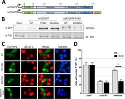
The Methyltransferase-Like Domain of Chikungunya Virus nsP2 Inhibits the Interferon Response by Promoting the Nuclear Export of STAT1.
Interferon Gamma Induces Reversible Metabolic Reprogramming of M1 Macrophages to Sustain Cell Viability and Pro-Inflammatory Activity.
Granzyme B PET Imaging as a Predictive Biomarker of Immunotherapy Response.
Deater M, Tamhankar M, Lloyd RE
PLoS pathogens 2022 Jan;18(1):e1010249
PLoS pathogens 2022 Jan;18(1):e1010249
SARS-CoV-2 nsp12 attenuates type I interferon production by inhibiting IRF3 nuclear translocation.
Wang W, Zhou Z, Xiao X, Tian Z, Dong X, Wang C, Li L, Ren L, Lei X, Xiang Z, Wang J
Cellular & molecular immunology 2021 Apr;18(4):945-953
Cellular & molecular immunology 2021 Apr;18(4):945-953
Activation and evasion of type I interferon responses by SARS-CoV-2.
Lei X, Dong X, Ma R, Wang W, Xiao X, Tian Z, Wang C, Wang Y, Li L, Ren L, Guo F, Zhao Z, Zhou Z, Xiang Z, Wang J
Nature communications 2020 Jul 30;11(1):3810
Nature communications 2020 Jul 30;11(1):3810
Suppression of Stromal Interferon Signaling by Human Papillomavirus 16.
Raikhy G, Woodby BL, Scott ML, Shin G, Myers JE, Scott RS, Bodily JM
Journal of virology 2019 Oct 1;93(19)
Journal of virology 2019 Oct 1;93(19)
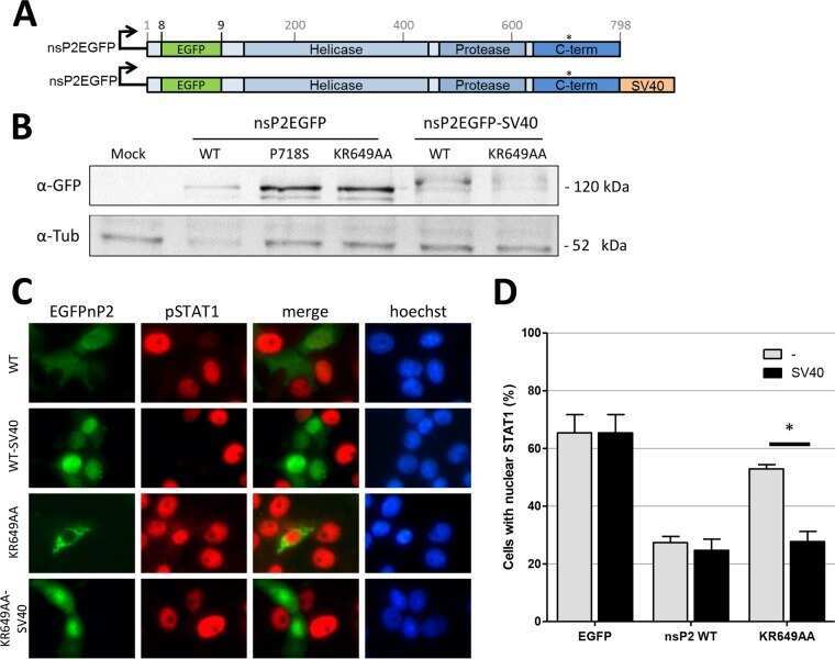
The Methyltransferase-Like Domain of Chikungunya Virus nsP2 Inhibits the Interferon Response by Promoting the Nuclear Export of STAT1.
Göertz GP, McNally KL, Robertson SJ, Best SM, Pijlman GP, Fros JJ
Journal of virology 2018 Sep 1;92(17)
Journal of virology 2018 Sep 1;92(17)
Interferon Gamma Induces Reversible Metabolic Reprogramming of M1 Macrophages to Sustain Cell Viability and Pro-Inflammatory Activity.
Wang F, Zhang S, Jeon R, Vuckovic I, Jiang X, Lerman A, Folmes CD, Dzeja PD, Herrmann J
EBioMedicine 2018 Apr;30:303-316
EBioMedicine 2018 Apr;30:303-316
Granzyme B PET Imaging as a Predictive Biomarker of Immunotherapy Response.
Larimer BM, Wehrenberg-Klee E, Dubois F, Mehta A, Kalomeris T, Flaherty K, Boland G, Mahmood U
Cancer research 2017 May 1;77(9):2318-2327
Cancer research 2017 May 1;77(9):2318-2327
No comments: Submit comment
Supportive validation
- Submitted by
- Invitrogen Antibodies (provider)
- Main image

- Experimental details
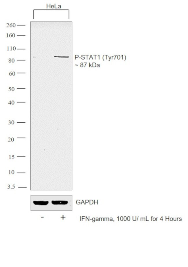
- Western blot analysis of Phospho-STAT1 pTyr701 in unstimulated HeLa lysates (lane 1) or lysates stimulated with 100 ng/mL IFN-gamma (lanes 2-4) using a Phospho-STAT1 pTyr701 recombinant rabbit monoclonal antibody (Product # 700349) at a dilution of 0.5 µg/mL.
- Submitted by
- Invitrogen Antibodies (provider)
- Main image

- Experimental details
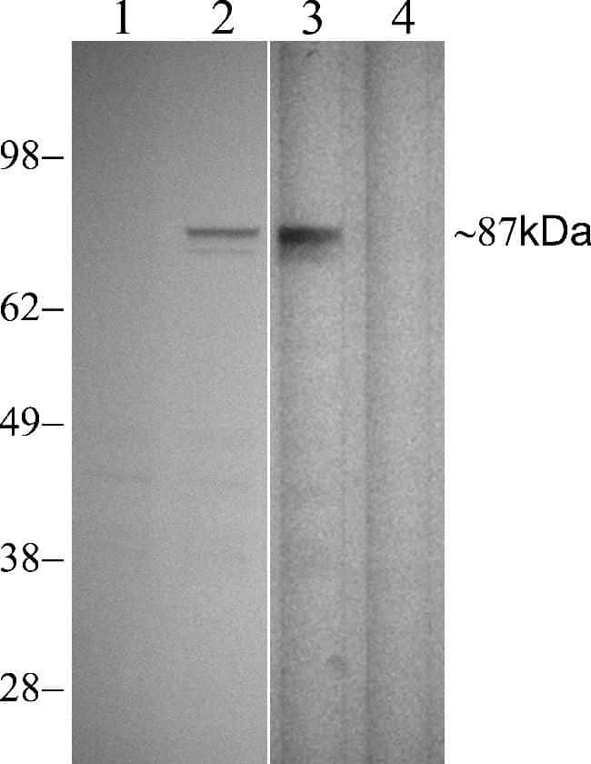
- Western blot was performed using Anti-Phospho-STAT1 (Tyr701) Rabbit Monoclonal Antibody (Product # 700349) and a 87 kDa band corresponding to STAT1 was observed in HeLa cells upon IFN gamma treatment. Whole cell extracts (30 µg lysate) of HeLa (Lane 1), HeLa treated with IFN-gamma (1000u/mL for 4 Hours) (Lane 2) were electrophoresed using Novex® NuPAGE® 4-12 % Bis-Tris gel (Product # NP0322BOX). Resolved proteins were then transferred onto a nitrocellulose membrane (Product # IB23001) by iBlot® 2 Dry Blotting System (Product # IB21001). The blot was probed with the primary antibody (1 µg/mL) and detected by chemiluminescence Goat Anti-Rabbit IgG Secondary Antibody, HRP conjugate (Product # A27036, 1:4000 dilution) using the iBright FL 1000 (Product # A32752). Chemiluminescent detection was performed using Novex® ECL Chemiluminescent Substrate Reagent Kit (Product # WP20005).
Supportive validation
- Submitted by
- Invitrogen Antibodies (provider)
- Main image

- Experimental details
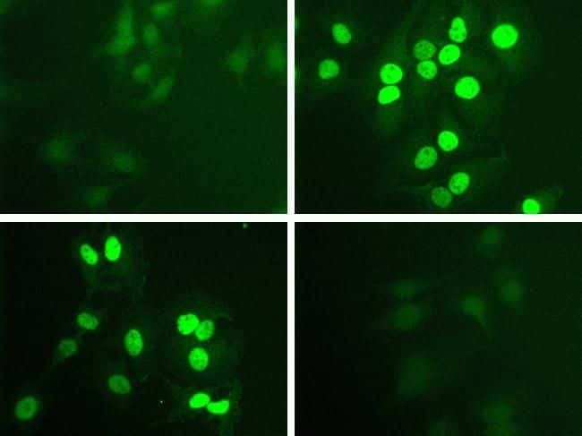
- Immunofluorescent analysis of Phospho-STAT1 pTyr701 in HeLa cells stimulated with 100 ng/mL IFN-gamma (top right) or unstimulated (top left) using a Phospho-STAT1 pTyr701 recombinant rabbit monoclonal antibody (Product # 700349) at a dilution of 1 µg/mL preincubated with the phosphopeptide used as an immunogen (bottom right) or with non-phosphopeptide (bottom left), followed by detection using an Alexa Fluor 488-conjugated goat anti-rabbit secondary antibody at a dilution of 1:1000.
Supportive validation
- Submitted by
- Invitrogen Antibodies (provider)
- Main image
- Experimental details
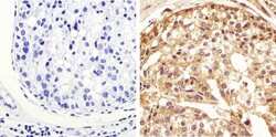
- Immunohistochemistry analysis of STAT1 (pY701) showing staining in the cytoplasm and nucleus of paraffin-embedded mouse human breast carcinoma (right) compared to a negative control without primary antibody (left). To expose target proteins, antigen retrieval was performed using 10mM sodium citrate (pH 6.0), microwaved for 8-15 min. Following antigen retrieval, tissues were blocked in 3% H2O2-methanol for 15 min at room temperature, washed with ddH2O and PBS, and then probed with a STAT1 (pY701) Monoclonal Antibody (clone 15H13L67), Recombinant (Product # 700349) diluted in 3% BSA-PBS at a dilution of 1:50 overnight at 4°C in a humidified chamber. Tissues were washed extensively in PBST and detection was performed using an HRP-conjugated secondary antibody followed by colorimetric detection using a DAB kit. Tissues were counterstained with hematoxylin and dehydrated with ethanol and xylene to prep for mounting.
- Submitted by
- Invitrogen Antibodies (provider)
- Main image

- Experimental details
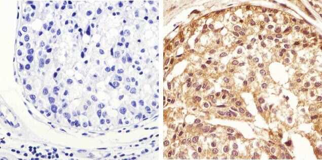
- Immunohistochemistry analysis of STAT1 (pY701) showing staining in the cytoplasm and nucleus of paraffin-embedded human cervical carcinoma (right) compared to a negative control without primary antibody (left). To expose target proteins, antigen retrieval was performed using 10mM sodium citrate (pH 6.0), microwaved for 8-15 min. Following antigen retrieval, tissues were blocked in 3% H2O2-methanol for 15 min at room temperature, washed with ddH2O and PBS, and then probed with a STAT1 (pY701) Monoclonal Antibody (clone 15H13L67), Recombinant (Product # 700349) diluted in 3% BSA-PBS at a dilution of 1:20 overnight at 4°C in a humidified chamber. Tissues were washed extensively in PBST and detection was performed using an HRP-conjugated secondary antibody followed by colorimetric detection using a DAB kit. Tissues were counterstained with hematoxylin and dehydrated with ethanol and xylene to prep for mounting.
Supportive validation
- Submitted by
- Invitrogen Antibodies (provider)
- Main image
- Experimental details
- NULL
- Submitted by
- Invitrogen Antibodies (provider)
- Main image

- Experimental details
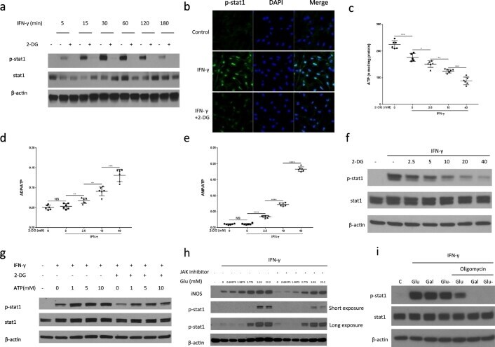
- Fig. 3 2-DG reduces IFN-gamma-induced JAK-STAT1 pathway activation by reducing ATP production. (a) Immunoblot analysis of specified proteins in BMDMs as a function of time of IFN-gamma stimulation +-1 hour pre-treatment with 2-DG (10 mM). Data are representative of two independent experiments. (b) Immunofluorescence staining of p-STAT-1 in BMDMs after incubation with IFN-gamma for 30 min +- 1 hour pre-treatment with 2-DG (10 mM). Data are representative of two independent experiments. (c) to (e) ATP production, ADP/ATP, AMP/ATP ratio of Raw264.7 cells, measured by HPLC after stimulation with IFN-gamma for 30 min +- 1 hour pre-treatment with 2-DG (10 mM). *p < 0.05, **p < 0.01, ***p < 0.001, ****p < 0.0001, NS: no significant difference. Data are represented as mean +- sem (n = 6). (f) Immunoblot analysis of specified proteins in Raw264.7 cells after stimulation with IFN-gamma for 30 min +- 1 hour pre-treatment with indicated concentrations of 2-DG. Data are representative of two independent experiments, (g) Immunoblot analysis of specified proteins in Raw264.7 cells after permeabilization with SLO (100 ng/ml), incubation with ATP (0-10 mM) for 15 min and 1-hour stimulation with IFN-gamma (+-10 mM 2-DG). Data are representative of two independent experiments. (h) Immunoblot analysis of specified proteins in Raw264.7 cells after stimulation with IFN-gamma for 9 h in medium containing different concentrations of glucose +-1 hour pre-treatment with JAK inhibitor I (100 nM). Data
- Submitted by
- Invitrogen Antibodies (provider)
- Main image
- Experimental details
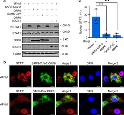
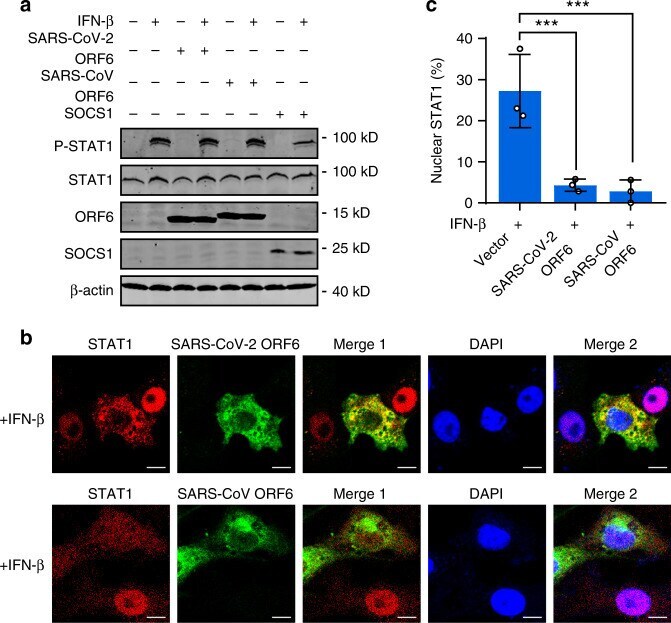
- Fig. 6 ORF6 inhibits STAT1 nuclear translocation but not phosphorylation. a Effect of SARS-CoV ORF6 and SARS-CoV-2 ORF6 on IFN-beta-induced phosphorylation of STAT1. HEK293T cells were transfected with a control plasmid or with plasmids expressing SARS-CoV ORF6, SARS-CoV-2 ORF6, or SOCS1. At 24 h after transfection, cells were left untreated or treated with 1000 U/mL IFN-beta for 30 min. The phosphorylation of STAT1 was detected by Western blot analyses. b Effect of SARS-CoV ORF6 and SARS-CoV-2 ORF6 on IFN-beta-induced nuclear translocation of STAT1. Vero cells were transfected with plasmids expressing SARS-CoV ORF6 and SARS-CoV-2 ORF6. At 24 h after transfection, cells were treated with 1000 U/mL IFN-beta for 30 min and stained with indicated antibodies. Merge 1 and Merge 2 indicate the merged red and green channels and the merged red, green, and blue channels, respectively. Scale bar, 10 mum. c Quantitation of the nuclear translocation of STAT1. All experiments were done at least twice, and one representative is shown. Error bars indicate SD of technical triplicates. *** P < 0.001, two-tailed Student's t -test. Source data are provided as a Source Data file.
- Submitted by
- Invitrogen Antibodies (provider)
- Main image

- Experimental details
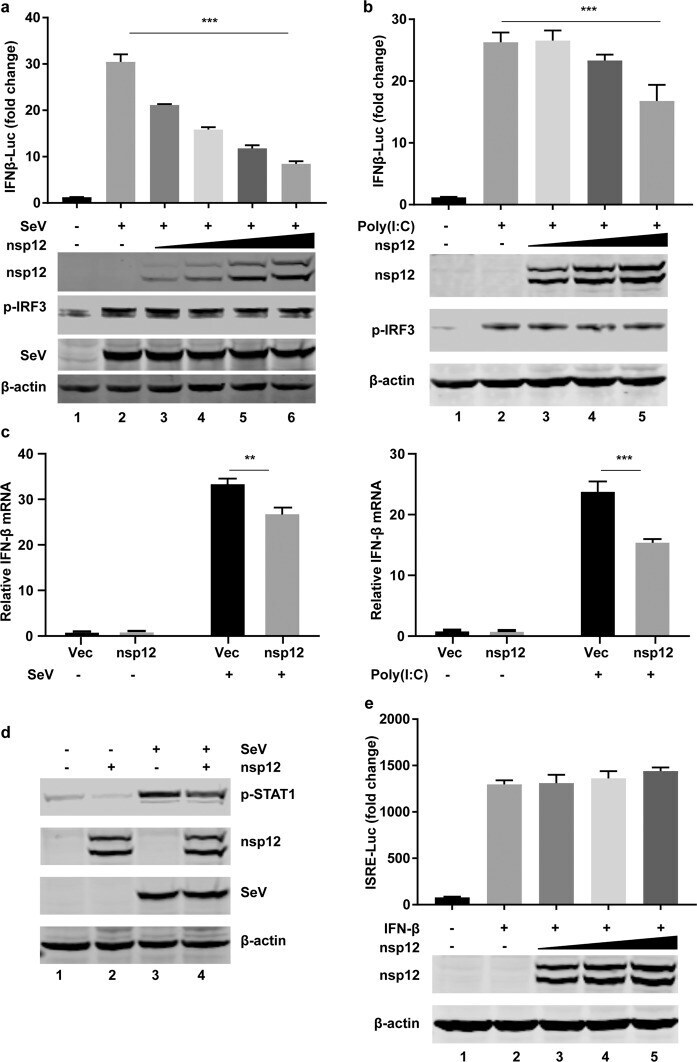
- Fig. 1 SARS-CoV-2 nsp12 attenuates viral RNA-related type I interferon responses. a Effects of nsp12 on SeV-induced IFN-beta promoter activation. HEK293T cells were transfected with an IFN-beta reporter plasmid along with a control plasmid or with increasing amounts of plasmids expressing nsp12. The cells were infected with SeV for 12 h and assayed for luciferase activity. The indicated protein expression levels were analyzed by western blotting. p-IRF3 was used to assess the stimulation after SeV infection. b Effects of nsp12 on high-molecular weight poly(I:C)-induced IFN-beta promoter activation. HEK293T cells were transfected with an IFN-beta reporter plasmid along with a control plasmid or with increasing amounts of plasmids expressing nsp12. The cells were transfected with high-molecular weight poly(I:C) for 12 h and assayed for luciferase activity. The indicated protein expression levels were analyzed by western blotting. p-IRF3 was used to assess the stimulation of poly I:C treatment. c Effect of nsp12 on endogenous IFN-beta mRNA expression induced by SeV (left) and high-molecular weight poly(I:C) (right). HEK293T cells were transfected with a control plasmid or a plasmid expressing nsp12. After 24 h, cells were infected with SeV for 8 h or transfected with high-molecular weight poly(I:C) for 6 h. Total RNA was extracted, and the expression of IFN-beta was detected by real-time RT-PCR. d Effect of nsp12 on endogenous p-STAT1 induced by SeV. HEK293T cells were transfect