 Explore
 Validate
 Learn
Learn Western blot
 ELISA
ELISAAntibody data
- Antibody Data
- Antigen structure
- References [21]
- Comments [0]
- Validations
- Western blot [3]
- Immunohistochemistry [2]
- Chromatin Immunoprecipitation [1]
- Other assay [8]
Submit
Validation data
Reference
Comment
Report error
- Product number
- 33-3400 - Provider product page

- Provider
- Invitrogen Antibodies
- Product name
- Phospho-STAT1 (Tyr701) Monoclonal Antibody (ST1P-11A5)
- Antibody type
- Monoclonal
- Antigen
- Synthetic peptide
- Description
- This antibody reacts specifically with the tyrosine-701 phosphorylated form of STAT1 and does not exhibit appreciable cross-reactivity with corresponding tyrosine phosphorylated forms of other STAT proteins or with other endogenous phosphotyrosine-containing proteins. To confirm the exclusive recognition of tyrosine phosphorylated STAT1, Western blots were carried out on lysates of 293 cells transfected with a STAT1 expression vector together with a wild type or kinase “dead” JAK1 expression vector. In addition, recognition of endogenous tyrosine phosphorylated STAT1 was confirmed on Western blots with cell lysates derived from serum starved or EGF-stimulated A431 cells and on Western blots with IFNa-stimulated mouse embryo fibroblast lysates.
- Reactivity
- Human, Mouse
- Host
- Mouse
- Isotype
- IgG
- Antibody clone number
- ST1P-11A5
- Vial size
- 50 µg
- Concentration
- 0.5 mg/mL
- Storage
- -20°C
Submitted references PBRM1 loss defines a nonimmunogenic tumor phenotype associated with checkpoint inhibitor resistance in renal carcinoma.
Signal transducer and activator of transcription (STAT) 1 and STAT3 are expressed in the human ovary and have Janus kinase 1-independent functions in the COV434 human granulosa cell line.
Analysis of interleukin-20 receptor complexes in trabecular meshwork cells and effects of cytokine signaling in anterior segment perfusion culture.
TWEAK blockade decreases atherosclerotic lesion size and progression through suppression of STAT1 signaling in diabetic mice.
Vaccinia Virus Protein C6 Inhibits Type I IFN Signalling in the Nucleus and Binds to the Transactivation Domain of STAT2.
PDGFRβ signalling regulates local inflammation and synergizes with hypercholesterolaemia to promote atherosclerosis.
STAT3 induction of miR-146b forms a feedback loop to inhibit the NF-κB to IL-6 signaling axis and STAT3-driven cancer phenotypes.
Chemokine gene silencing in decidual stromal cells limits T cell access to the maternal-fetal interface.
PDlim2 selectively interacts with the PDZ binding motif of highly pathogenic avian H5N1 influenza A virus NS1.
The JAK2/STAT3 signaling pathway is required for growth of CD44⁺CD24⁻ stem cell-like breast cancer cells in human tumors.
IL-27 directly restrains lung tumorigenicity by suppressing cyclooxygenase-2-mediated activities.
Functional genomics highlights differential induction of antiviral pathways in the lungs of SARS-CoV-infected macaques.
Identification of human STAT5-dependent gene regulatory elements based on interspecies homology.
Double-stranded RNAs from the helminth parasite Schistosoma activate TLR3 in dendritic cells.
Signal regulatory protein alpha ligation induces macrophage nitric oxide production through JAK/STAT- and phosphatidylinositol 3-kinase/Rac1/NAPDH oxidase/H2O2-dependent pathways.
The protective effect of moderate hypothermia during intestinal ischemia-reperfusion is associated with modification of hepatic transcription factor activation.
Mechanisms of oncogenic KIT signal transduction in primary gastrointestinal stromal tumors (GISTs).
Rapamycin inhibits the interleukin 10 signal transduction pathway and the growth of Epstein Barr virus B-cell lymphomas.
Constitutive activation of Jak/STAT proteins in Epstein-Barr virus-infected B-cell lines from patients with posttransplant lymphoproliferative disorder.
Constitutive activation of Jak/STAT proteins in Epstein-Barr virus-infected B-cell lines from patients with posttransplant lymphoproliferative disorder.
Activation of Stat3 in v-Src-transformed fibroblasts requires cooperation of Jak1 kinase activity.
Liu XD, Kong W, Peterson CB, McGrail DJ, Hoang A, Zhang X, Lam T, Pilie PG, Zhu H, Beckermann KE, Haake SM, Isgandrova S, Martinez-Moczygemba M, Sahni N, Tannir NM, Lin SY, Rathmell WK, Jonasch E
Nature communications 2020 May 1;11(1):2135
Nature communications 2020 May 1;11(1):2135
Signal transducer and activator of transcription (STAT) 1 and STAT3 are expressed in the human ovary and have Janus kinase 1-independent functions in the COV434 human granulosa cell line.
Frost ER, Ford EA, Peters AE, Reed NL, McLaughlin EA, Baker MA, Lovell-Badge R, Sutherland JM
Reproduction, fertility, and development 2020 Aug;32(12):1027-1039
Reproduction, fertility, and development 2020 Aug;32(12):1027-1039
Analysis of interleukin-20 receptor complexes in trabecular meshwork cells and effects of cytokine signaling in anterior segment perfusion culture.
Keller KE, Yang YF, Sun YY, Walter MR, Wirtz MK
Molecular vision 2019;25:266-282
Molecular vision 2019;25:266-282
TWEAK blockade decreases atherosclerotic lesion size and progression through suppression of STAT1 signaling in diabetic mice.
Fernández-Laso V, Sastre C, Méndez-Barbero N, Egido J, Martín-Ventura JL, Gómez-Guerrero C, Blanco-Colio LM
Scientific reports 2017 Apr 27;7:46679
Scientific reports 2017 Apr 27;7:46679
Vaccinia Virus Protein C6 Inhibits Type I IFN Signalling in the Nucleus and Binds to the Transactivation Domain of STAT2.
Stuart JH, Sumner RP, Lu Y, Snowden JS, Smith GL
PLoS pathogens 2016 Dec;12(12):e1005955
PLoS pathogens 2016 Dec;12(12):e1005955
PDGFRβ signalling regulates local inflammation and synergizes with hypercholesterolaemia to promote atherosclerosis.
He C, Medley SC, Hu T, Hinsdale ME, Lupu F, Virmani R, Olson LE
Nature communications 2015 Jul 17;6:7770
Nature communications 2015 Jul 17;6:7770
STAT3 induction of miR-146b forms a feedback loop to inhibit the NF-κB to IL-6 signaling axis and STAT3-driven cancer phenotypes.
Xiang M, Birkbak NJ, Vafaizadeh V, Walker SR, Yeh JE, Liu S, Kroll Y, Boldin M, Taganov K, Groner B, Richardson AL, Frank DA
Science signaling 2014 Jan 28;7(310):ra11
Science signaling 2014 Jan 28;7(310):ra11
Chemokine gene silencing in decidual stromal cells limits T cell access to the maternal-fetal interface.
Nancy P, Tagliani E, Tay CS, Asp P, Levy DE, Erlebacher A
Science (New York, N.Y.) 2012 Jun 8;336(6086):1317-21
Science (New York, N.Y.) 2012 Jun 8;336(6086):1317-21
PDlim2 selectively interacts with the PDZ binding motif of highly pathogenic avian H5N1 influenza A virus NS1.
Yu J, Li X, Wang Y, Li B, Li H, Li Y, Zhou W, Zhang C, Wang Y, Rao Z, Bartlam M, Cao Y
PloS one 2011;6(5):e19511
PloS one 2011;6(5):e19511
The JAK2/STAT3 signaling pathway is required for growth of CD44⁺CD24⁻ stem cell-like breast cancer cells in human tumors.
Marotta LL, Almendro V, Marusyk A, Shipitsin M, Schemme J, Walker SR, Bloushtain-Qimron N, Kim JJ, Choudhury SA, Maruyama R, Wu Z, Gönen M, Mulvey LA, Bessarabova MO, Huh SJ, Silver SJ, Kim SY, Park SY, Lee HE, Anderson KS, Richardson AL, Nikolskaya T, Nikolsky Y, Liu XS, Root DE, Hahn WC, Frank DA, Polyak K
The Journal of clinical investigation 2011 Jul;121(7):2723-35
The Journal of clinical investigation 2011 Jul;121(7):2723-35
IL-27 directly restrains lung tumorigenicity by suppressing cyclooxygenase-2-mediated activities.
Ho MY, Leu SJ, Sun GH, Tao MH, Tang SJ, Sun KH
Journal of immunology (Baltimore, Md. : 1950) 2009 Nov 15;183(10):6217-26
Journal of immunology (Baltimore, Md. : 1950) 2009 Nov 15;183(10):6217-26
Functional genomics highlights differential induction of antiviral pathways in the lungs of SARS-CoV-infected macaques.
de Lang A, Baas T, Teal T, Leijten LM, Rain B, Osterhaus AD, Haagmans BL, Katze MG
PLoS pathogens 2007 Aug 10;3(8):e112
PLoS pathogens 2007 Aug 10;3(8):e112
Identification of human STAT5-dependent gene regulatory elements based on interspecies homology.
Nelson EA, Walker SR, Li W, Liu XS, Frank DA
The Journal of biological chemistry 2006 Sep 8;281(36):26216-24
The Journal of biological chemistry 2006 Sep 8;281(36):26216-24
Double-stranded RNAs from the helminth parasite Schistosoma activate TLR3 in dendritic cells.
Aksoy E, Zouain CS, Vanhoutte F, Fontaine J, Pavelka N, Thieblemont N, Willems F, Ricciardi-Castagnoli P, Goldman M, Capron M, Ryffel B, Trottein F
The Journal of biological chemistry 2005 Jan 7;280(1):277-83
The Journal of biological chemistry 2005 Jan 7;280(1):277-83
Signal regulatory protein alpha ligation induces macrophage nitric oxide production through JAK/STAT- and phosphatidylinositol 3-kinase/Rac1/NAPDH oxidase/H2O2-dependent pathways.
Alblas J, Honing H, de Lavalette CR, Brown MH, Dijkstra CD, van den Berg TK
Molecular and cellular biology 2005 Aug;25(16):7181-92
Molecular and cellular biology 2005 Aug;25(16):7181-92
The protective effect of moderate hypothermia during intestinal ischemia-reperfusion is associated with modification of hepatic transcription factor activation.
Parkinson EJ, Townsend PA, Stephanou A, Latchman DS, Eaton S, Pierro A
Journal of pediatric surgery 2004 May;39(5):696-701
Journal of pediatric surgery 2004 May;39(5):696-701
Mechanisms of oncogenic KIT signal transduction in primary gastrointestinal stromal tumors (GISTs).
Duensing A, Medeiros F, McConarty B, Joseph NE, Panigrahy D, Singer S, Fletcher CD, Demetri GD, Fletcher JA
Oncogene 2004 May 13;23(22):3999-4006
Oncogene 2004 May 13;23(22):3999-4006
Rapamycin inhibits the interleukin 10 signal transduction pathway and the growth of Epstein Barr virus B-cell lymphomas.
Nepomuceno RR, Balatoni CE, Natkunam Y, Snow AL, Krams SM, Martinez OM
Cancer research 2003 Aug 1;63(15):4472-80
Cancer research 2003 Aug 1;63(15):4472-80
Constitutive activation of Jak/STAT proteins in Epstein-Barr virus-infected B-cell lines from patients with posttransplant lymphoproliferative disorder.
Nepomuceno RR, Snow AL, Robert Beatty P, Krams SM, Martinez OM
Transplantation 2002 Aug 15;74(3):396-402
Transplantation 2002 Aug 15;74(3):396-402
Constitutive activation of Jak/STAT proteins in Epstein-Barr virus-infected B-cell lines from patients with posttransplant lymphoproliferative disorder.
Nepomuceno RR, Snow AL, Robert Beatty P, Krams SM, Martinez OM
Transplantation 2002 Aug 15;74(3):396-402
Transplantation 2002 Aug 15;74(3):396-402
Activation of Stat3 in v-Src-transformed fibroblasts requires cooperation of Jak1 kinase activity.
Zhang Y, Turkson J, Carter-Su C, Smithgall T, Levitzki A, Kraker A, Krolewski JJ, Medveczky P, Jove R
The Journal of biological chemistry 2000 Aug 11;275(32):24935-44
The Journal of biological chemistry 2000 Aug 11;275(32):24935-44
No comments: Submit comment
Supportive validation
- Submitted by
- Invitrogen Antibodies (provider)
- Main image

- Experimental details
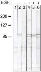
- Western blot analysis of phospho-STAT1 expression in unstimulated (-) or EGF-stimulated (+) A431 cells using Rabbit anti-phospho-STAT1 (Lane 1 & Lane 4), Rabbit anti-STAT1 (Lane 2 & Lane 5), Mouse anti-phospho-STAT1 (Lane 3 & Lane 6) (Product # 33-3400).
- Submitted by
- Invitrogen Antibodies (provider)
- Main image

- Experimental details
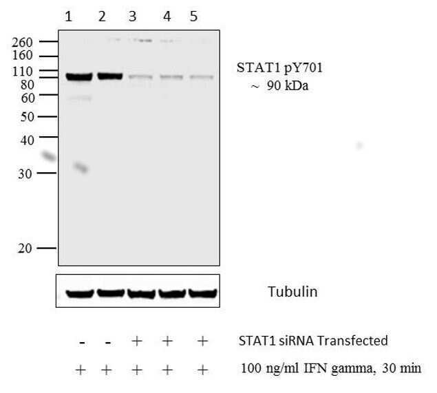
- Western blot analysis of STAT1 (pT701) was performed with 20 µg of HeLa cells treated for 30 minutes with 100 ng/mL of IFN gamma (lane1), HeLa transfected with 0 nM, 20 nM, 50, 100 nM Silencer® Select STAT1 siRNA (s277) (lane 2, 3, 4, 5 respectively) and treated for 30 minutes with 100 ng/mL of IFN gamma, HeLa (lane6) cell lysate using Novex®NuPAGE® 10 % Bis-Tris gel (Product # NP0302BOX), XCell SureLock Electrophoresis System (Product # EI0002), Novex® Sharp Pre-Stained Protein Standard (LC5800). Proteins were transferred using Pierce G2 Fast Blotter (Product # 62287) to a nitrocellulose membrane and blocked with 5% skim milk for 1 hour at room temperature. STAT1 (pT701) was detected at ~ 90 kDa using STAT1 (pT701) Mouse Monoclonal Antibody (Product # 33-3400) at 2 µg/mL in 5% skim milk at 4°C overnight on a rocking platform. Goat Anti-Mouse - HRP Secondary Antibody (Product # 62-6520) at 1:4000 dilution was used and chemiluminescent detection was performed using Pierce™ ECL Western Blotting Substrate (Product # 32106).STAT1 (pT701) Antibody (Product # 33-3400) confirms silencing of STAT1 expression.
- Submitted by
- Invitrogen Antibodies (provider)
- Main image

- Experimental details
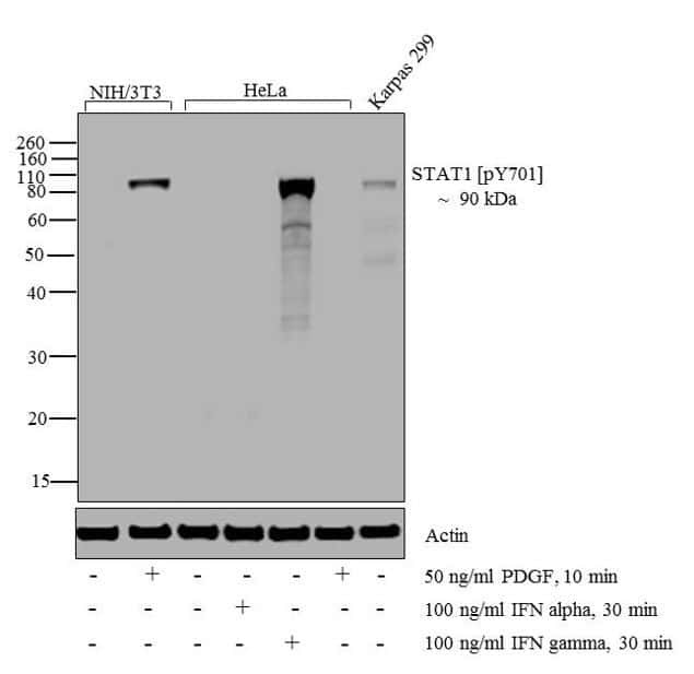
- Western blot analysis of STAT1 (pT701) was performed by loading 20 µg of NIH/3T3 (lane1), NIH/3T3 treated for 10 minutes with 50 ng/mL of PDGF (lane2), HeLa (lane3), HeLa treated for 30 minutes with 100 ng/mL of IFN alpha (lane4), HeLa treated for 30 minutes with 100 ng/mL of IFN gamma (lane5), HeLa treated for 10 minutes with 50 ng/mL of PDGF (lane6) and Karpas 299 (lane7) cell lysate using Novex®NuPAGE® 10 % Bis-Tris gel (Product # NP0302BOX), XCell SureLock Electrophoresis System (Product # EI0002), Novex® Sharp Pre-Stained Protein Standard (LC5800), and iBlot® Dry Blotting System (IB21001). Proteins were transferred to a nitrocellulose membrane and blocked with 5% skim milk at 4°C overnight. STAT1 (pT701) was detected at ~ 90 kDa using STAT1 (pT701) Mouse Monoclonal Antibody (Product # 33-3400) at 0.5-1 µg/mL in 5% skim milk for 3 hour at room temperature on a rocking platform. Goat Anti-Mouse - HRP Secondary Antibody (Product # 62-6520) at 1:4000 dilution was used and chemiluminescent detection was performed using Pierce™ ECL Western Blotting Substrate (Product # 32106).
Supportive validation
- Submitted by
- Invitrogen Antibodies (provider)
- Main image

- Experimental details
- Immunohistochemistry analysis of Phospho-STAT1 (pTyr701) showing staining in the nucleus and cytoplasm of paraffin-embedded human cervical carcinoma tissue (right) compared to a negative control without primary antibody (left). To expose target proteins, antigen retrieval was performed using 10mM sodium citrate (pH 6.0), microwaved for 8-15 min. Following antigen retrieval, tissues were blocked in 3% H2O2-methanol for 15 min at room temperature, washed with ddH2O and PBS, and then probed with a Phospho-STAT1 pTyr701monoclonal antibody (Product # 33-3400) diluted in 3% BSA-PBS at a dilution of 1:20 overnight at 4°C in a humidified chamber. Tissues were washed extensively in PBST and detection was performed using an HRP-conjugated secondary antibody followed by colorimetric detection using a DAB kit. Tissues were counterstained with hematoxylin and dehydrated with ethanol and xylene to prep for mounting.

- Submitted by
- Invitrogen Antibodies (provider)
- Main image

- Experimental details
- Immunohistochemistry analysis of STAT1 (pY701) showing staining in the cytoplasm and nucleus of paraffin-embedded human breast carcinoma tissue (right) compared to a negative control without primary antibody (left). To expose target proteins, antigen retrieval was performed using 10mM sodium citrate (pH 6.0), microwaved for 8-15 min. Following antigen retrieval, tissues were blocked in 3% H2O2-methanol for 15 min at room temperature, washed with ddH2O and PBS, and then probed with a STAT1 (pY701) monoclonal antibody (Product # 33-3400) diluted in 3% BSA-PBS at a dilution of 1:20 overnight at 4ºC in a humidified chamber. Tissues were washed extensively in PBST and detection was performed using an HRP-conjugated secondary antibody followed by colorimetric detection using a DAB kit. Tissues were counterstained with hematoxylin and dehydrated with ethanol and xylene to prep for mounting.

Supportive validation
- Submitted by
- Invitrogen Antibodies (provider)
- Main image

- Experimental details
- ChIP- qPCR analysis of STAT1 (pTyr701) was performed with 5 µg of the STAT1 (pTyr701) Mouse monoclonal antibody (ST1P-11A5) (Product # 33-3400) on sheared chromatin from 2 million HeLa cells treated with 50 ng/mL of IFN Gamma for one hour using the MAGnify Chromatin Immunoprecipitation System (Product # 49-2024). Normal Mouse IgG was used as a negative IP control. The purified DNA from each ChIP sample was analyzed by StepOnePlus Real-Time PCR System (Product # 4376600) with primers for the promoter of active GLS1 gene, used as positive control target, and the inactive SAT2, used as negative control target. Data is presented as fold enrichment of the antibody signal versus the negative control IgG using the comparative CT method.

Supportive validation
- Submitted by
- Invitrogen Antibodies (provider)
- Main image

- Experimental details
- NULL

- Submitted by
- Invitrogen Antibodies (provider)
- Main image

- Experimental details
- Figure 6 Effects of the PDlim2 and HN12-NS1 interaction on NF-kappaB activity or STAT1 phosphorylation. (A) HeLa cells was transfected with the reporter plasmid pNF-kappaB-luc and ptk-RL, together with the indicated plasmids. After 24-hour post-transfection, cells were stimulated by LPS (10 ug/ml) for 5 hours. Cells were harvested and firefly and renilla fluorescence unit were measured by luminomitor. The relative luciferase activity (Y-axis) = the unit of firefly luciferase activity / the unit of Renilla luciferase activity (to indicat transfection efficiency, and is presented as the mean calculated from three independent experiments. The expression of NS1 and PDlim2 were analyzed by Western blot with indicated antibodies (lower panels). (B) HeLa cells were transfected with the combination of the plasmids as indicated at the top of the figure and treated by IFN-alpha at 50 U/ml for 4 hours. The cell lysates were analyzed by SDS-PAGE and Western blot, with antibodies specific for phosphorylated STAT1, STAT1, HA-tag, Flag-tag, and GAPDH as loading control.

- Submitted by
- Invitrogen Antibodies (provider)
- Main image

- Experimental details
- Figure 7 PDGF signaling activates STAT1 in VSMCs (a) Tyrosine phosphorylation of STAT1, STAT3, and STAT5 in cultured VSMCs from PDGFRbeta Sm22D849V (beta-D849V) mutant and Sm22alpha-Cre (Wt) control mice was determined by Western blot. As a positive control, VSMCs were treated with 10ng ml -1 mouse IFN-gamma for 30 min. (b) Phosphorylation of PDGFRbeta and STAT1 in control VSMCs after serum starvation and treatment with 10 ng ml -1 PDGF-BB for 15 minutes was determined by Western blot. PDGFRbeta was immunoprecipitated from cell lysate and then phosphorylation was detected with anti-phosphotyrosine antibody. Phospho-STAT1 was detected in cell lysate with p-Y701 antibody. (c, d) PDGFRbeta and STAT1 phosphorylation in PDGFRbeta Sm22D849V mutant and control VSMCs were analyzed by Western blotting after 16 hrs treatment with the PDGFR inhibitor 10 muM AG-1295. (e) STAT1 phosphorylation in PDGFRbeta Sm22D849V mutant and control VSMCs were analyzed by Western blotting after 16 hrs treatment with 10 muM AG-1295 followed by vehicle or 10ng ml -1 mouse IFN-gamma for 30 min. (f) Quantification of Western blots in (e) by densitometry shown as fold change over untreated Wt (lane 1). (g) STAT1 and PLCgamma1 phosphorylation in PDGFRbeta Sm22D849V mutant and control VSMCs were analyzed by Western blotting after 16 hrs treatment with the JAK1/JAK2 inhibitor Ruxolitinib (10 muM) followed by 10ng ml -1 mouse IFN-gamma for 30 minutes or 10 ng ml -1 PDGF-BB for 10 minutes. (h) Quantification of W

- Submitted by
- Invitrogen Antibodies (provider)
- Main image

- Experimental details
- Figure 8 STAT1 regulates chemokines and inflammation in PDGFRbeta Sm22D849V mice (a) Expression of total STAT1 and STAT1 p-Y701 in PDGFRbeta Sm22D849V (beta-D849V) mutant and Sm22alpha-Cre (Wt) control VSMCs after transfection with control or STAT1-blocking siRNA was analyzed by Western blotting. (b) Fold change in chemokine mRNA levels in cultured VSMCs from mutant mice was determined by quantitative RT-PCR. Results are shown as fold change in STAT1 siRNA compared to control siRNA, n=4 transfections per siRNA. (c) Expression of total STAT1 and p-Y701 STAT1 in PDGFRbeta Sm22D849V (beta-D849V) single mutant and STAT1 Sm22CKO PDGFRbeta Sm22D849V (beta-D849V,STAT1-cKO) double mutant VSMCs was analyzed by Western blotting. (d) Fold change in chemokine mRNA levels in Wt, STAT1 Sm22CKO (STAT1-cKO), PDGFRbeta Sm22D849V (beta-D849V) single mutant, and STAT1 Sm22CKO PDGFRbeta Sm22D849V (beta-D849V,STAT1-cKO) double mutant VSMCs was determined by quantitative RT-PCR. Results are shown as fold change compared to Wt, n=3 different VSMC isolates per genotype. (e) Quantification of CD45 + leukocytes in the whole aorta of single mutant and double mutant mice (all ApoE +/+ ) as measured by flow cytometry, n=3 mice per genotype. (f) Mature VSMC marker expression in the cross-sectioned thoracic aorta of 8-week old mice was determined by immunofluorescence, with DAPI as a nuclear counterstain. Representative images from three aortas of each genotype are shown. Scale bar, 50mum. All data were as

- Submitted by
- Invitrogen Antibodies (provider)
- Main image

- Experimental details
- Figure 2 PDGF signaling in VSMCs causes inflammation of the aorta (a) Flow cytometric analysis of cells in the aorta of PDGFRbeta Sm22D849V (beta-D849V) mutant mice and Sm22alpha-Cre (Wt) control mice after enzymatic digestion. Numerical data indicate the percentage of CD45 + leukocytes among the total cells analyzed. Representative plots from three aortas of each genotype are shown. (b, c) Quantification of CD45 + leukocytes in the whole aorta or the separated media and adventitia as measured by flow cytometry, n=3 mice per genotype for each type of analysis. (d) Distribution of leukocytes (red arrows) in the media and adventitia was determined by staining cross sections of the thoracic aorta with Masson's trichrome. Representative sections from 3 aortas of each genotype are shown. Scale bar, 100mum (e) Quantification of immune cell types in the aortic wall as measured by flow cytometry using fluorescent-conjugated antibodies: APC-CD3; APC.Cy7-CD19; PE-CD11c; PE.Cy7-NK1.1; PerCy5.5-Mac1, n=3 mice per genotype. (f) Phosphorylation of immunoprecipitated PDGFRbeta from cultured VSMCs was determined by Western blotting with anti-phosphotyrosine antibody. PDGFRbeta-D849V tyrosine kinase activity was inhibited by treatment with the PDGFR inhibitor Crenolanib (400ng ml -1 ) for 2 hours prior to cell lysis. Representative blot from 2 experiments is shown. (g) Quantification of CD45 + leukocytes in the whole aorta of PDGFRbeta Sm22D849V mutant mice was measured by flow cytometry

- Submitted by
- Invitrogen Antibodies (provider)
- Main image

- Experimental details
- Figure 7 Detection of IFN-beta and Phosphorylated STAT1 in Lung of SARS-CoV-Infected Macaques Using Immunohistochemistry (A) Lack of IFN-beta expression in lungs of mock-infected macaques (magnification 40x) and (B, C) expression of IFN-beta (red) in lungs of SARS-CoV-infected macaques at day 1 post infection (40x [B] and 100x [C], arrowheads). (D) Lack of phosphorylated STAT1 in lungs of mock-infected macaques (40x) and (E) abundant presence of phosphorylated STAT1 (brown) in lungs of SARS-CoV-infected macaques at day 1 post infection (40x, arrowheads). (F) No detection of phosphorylated STAT1 (brown) in SARS-CoV-infected cells (red) (40x, arrowheads).

- Submitted by
- Invitrogen Antibodies (provider)
- Main image

- Experimental details
- Fig 2 C6 does not inhibit the IFNalpha-induced phosphorylation of STAT1 or STAT2. HeLa cells stably expressing the proteins shown were stimulated with IFNalpha (1000 U/ml for 1 h). Cells were harvested and cell lysates subjected to SDS-PAGE and immunoblotting to assess levels of STAT1 and STAT2 phosphorylation ( A ). Samples were also immunoblotted for alpha tubulin and actin as controls. Positions of molecular mass markers are shown on the left of the figure. Quantification of triplicate samples of phosphorylated STAT1 ( B ) and phosphorylated STAT2 ( C ) proteins was performed using Odyssey software (LICOR) and are shown relative to a constant house-keeping gene. P

- Submitted by
- Invitrogen Antibodies (provider)
- Main image

- Experimental details
- Figure 4 Western immunoblot analysis of STAT phosphorylation. Western immunoblot analysis of signal transducer and activator of transcription (STAT)-1 Tyr701, STAT-3 Ser727, STAT-3 Tyr705, and STAT-5 Tyr694 phosphorylation in ( A ) normal human trabecular meshwork cells or ( B ) normal human dermal fibroblasts treated with the indicated interleukins for 15 min. Total STATs were used as loading controls. The arrow points to the upper phosphorylated band. C: Densitometry of the phosphorylated Ser727 STAT-3 band. Trabecular meshwork, n=6 technical replicates using four independent cell strains; dermal fibroblasts, n=3 technical replicates from three cell strains. * p
