Explore
 Validate
 Learn
Learn Western blot
 Immunohistochemistry
Antibody data
- Antibody Data
- Antigen structure
- References [2]
- Comments [0]
- Validations
- Immunohistochemistry [1]
Submit
Validation data
Reference
Comment
Report error
- Product number
- A00036-2 - Provider product page

- Provider
- Boster Biological Technology
- Product name
- Anti-STAT1 Antibody Picoband™
- Antibody type
- Polyclonal
- Description
- Rabbit IgG polyclonal antibody for Signal transducer and activator of transcription 1-alpha/beta(STAT1) detection. Tested with WB, IHC-P, IHC-F, ICC/IF, FCM in Human;Mouse;Monkey;Rat.
- Reactivity
- Human, Mouse, Rat, Simian
- Host
- Rabbit
- Vial size
- 100μg/vial
- Concentration
- Add 0.2ml of distilled water will yield a concentration of 500ug/ml.
- Storage
- At -20°C for one year. After reconstitution, at 4°C for one month. It can also be aliquoted and stored frozen at -20°C for a longer time. Avoid repeated freezing and thawing.
- Handling
- Add 0.2ml of distilled water will yield a concentration of 500ug/ml.
Submitted references Coinfection of Porcine Circovirus 2 and Pseudorabies Virus Enhances Immunosuppression and Inflammation through NF-κB, JAK/STAT, MAPK, and NLRP3 Pathways.
Death signal transduction induced by co-immobilized TNF-α plus IFN-γ and the development of polymeric anti-cancer drugs.
Li X, Chen S, Zhang L, Niu G, Zhang X, Yang L, Ji W, Ren L
International journal of molecular sciences 2022 Apr 18;23(8)
International journal of molecular sciences 2022 Apr 18;23(8)
Death signal transduction induced by co-immobilized TNF-α plus IFN-γ and the development of polymeric anti-cancer drugs.
Guan YQ, Li Z, Liu JM
Biomaterials 2010 Dec;31(34):9074-85
Biomaterials 2010 Dec;31(34):9074-85
No comments: Submit comment
Supportive validation
- Submitted by
- Boster Biological Technology (provider)
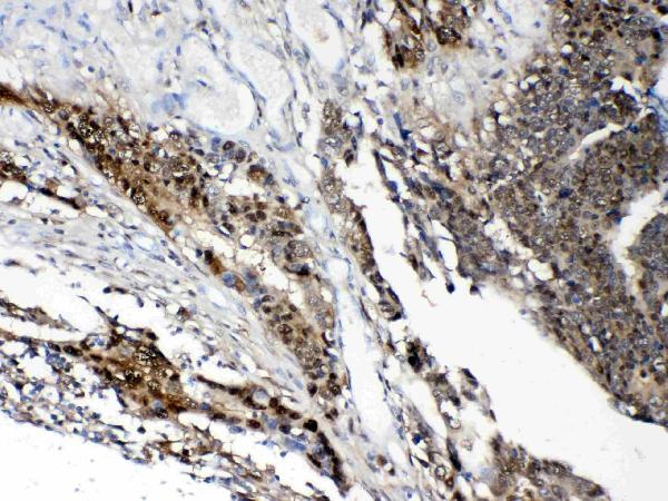
- Main image

- Experimental details
- IHC analysis of STAT1 using anti- STAT1 antibody (A00036-2).STAT1 was detected in paraffin-embedded section of human intestinal cancer tissues. Heat mediated antigen retrieval was performed in citrate buffer (pH6, epitope retrieval solution) for 20 mins. The tissue section was blocked with 10% goat serum. The tissue section was then incubated with 1μg/ml rabbit anti- STAT1 Antibody (A00036-2) overnight at 4°C. Biotinylated goat anti-rabbit IgG was used as secondary antibody and incubated for 30 minutes at 37°C. The tissue section was developed using Strepavidin-Biotin-Complex (SABC)(Catalog # SA1022) with DAB as the chromogen.
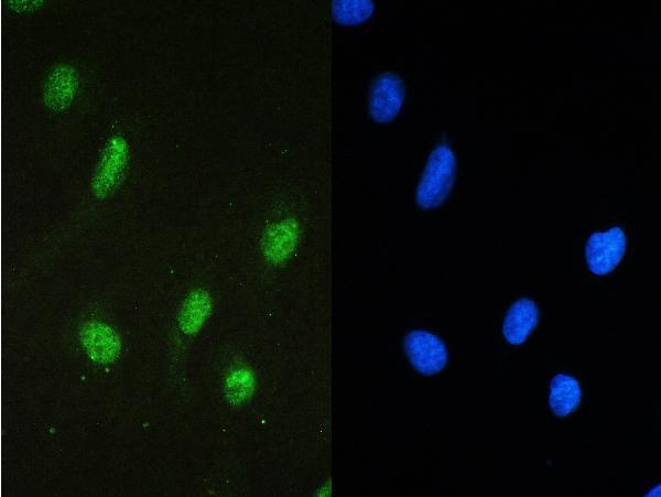
- Additional image