Explore
 Validate
 Learn
Learn Western blot
 Immunocytochemistry
Antibody data
- Antibody Data
- Antigen structure
- References [4]
- Comments [0]
- Validations
- Western blot [1]
Submit
Validation data
Reference
Comment
Report error
- Product number
- M00036-2 - Provider product page

- Provider
- Boster Biological Technology
- Product name
- Anti-STAT1 Antibody Picoband™ (monoclonal, 12C7)
- Antibody type
- Monoclonal
- Description
- Mouse IgG monoclonal antibody for STAT1 detection. Tested with WB, IHC-P, ICC/IF, FCM in Human;Monkey.
- Reactivity
- Human, Simian
- Host
- Mouse
- Isotype
- IgG
- Antibody clone number
- 12C7
- Vial size
- 100μg/vial
- Concentration
- 0.5-1mg/ml, actual concentration vary by lot. Use suggested dilution ratio to decide dilution procedure.
- Storage
- At -20°C for one year. After reconstitution, at 4°C for one month. It can also be aliquoted and stored frozen at -20°C for a longer time. Avoid repeated freezing and thawing.
- Handling
- Add 0.2ml of distilled water will yield a concentration of 500μg/ml.
Submitted references Blockage of Glyoxalase I Inhibits Colorectal Tumorigenesis and Tumor Growth via Upregulation of STAT1, p53, and Bax and Downregulation of c-Myc and Bcl-2.
Death signal transduction induced by co-immobilized TNF-α plus IFN-γ and the development of polymeric anti-cancer drugs.
Effects of acitretin on proliferative inhibition and RANTES production of HaCaT cells.
Hepatitis C virus non-structural 5A abrogates signal transducer and activator of transcription-1 nuclear translocation induced by IFN-alpha through dephosphorylation.
Chen Y, Fang L, Zhang J, Li G, Ma M, Li C, Lyu J, Meng QH
International journal of molecular sciences 2017 Mar 9;18(3)
International journal of molecular sciences 2017 Mar 9;18(3)
Death signal transduction induced by co-immobilized TNF-α plus IFN-γ and the development of polymeric anti-cancer drugs.
Guan YQ, Li Z, Liu JM
Biomaterials 2010 Dec;31(34):9074-85
Biomaterials 2010 Dec;31(34):9074-85
Effects of acitretin on proliferative inhibition and RANTES production of HaCaT cells.
Zhang M, Zhu L, Feng Y, Yang Y, Liu L, Ran Y
Archives of dermatological research 2008 Nov;300(10):575-81
Archives of dermatological research 2008 Nov;300(10):575-81
Hepatitis C virus non-structural 5A abrogates signal transducer and activator of transcription-1 nuclear translocation induced by IFN-alpha through dephosphorylation.
Gong GZ, Cao J, Jiang YF, Zhou Y, Liu B
World journal of gastroenterology 2007 Aug 14;13(30):4080-4
World journal of gastroenterology 2007 Aug 14;13(30):4080-4
No comments: Submit comment
Supportive validation
- Submitted by
- Boster Biological Technology (provider)
- Main image
- Experimental details
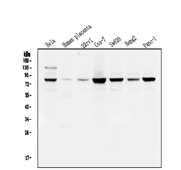
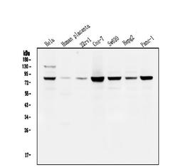
- Western blot analysis of STAT1 using anti-STAT1 antibody (M00036-2). Electrophoresis was performed on a 5-20% SDS-PAGE gel at 70V (Stacking gel) / 90V (Resolving gel) for 2-3 hours. The sample well of each lane was loaded with 50ug of sample under reducing conditions. Lane 1: human Hela whole cell lysates, Lane 2: human placenta tissue lysates, Lane 3: human 22RV1 whole cell lysates. Lane 4: monkey COS-7 whole cell lysates, Lane 5: human SW620 whole cell lysates, Lane 6: human HepG2 whole cell lysates, Lane 7: human PANC-1 whole cell lysates. After Electrophoresis, proteins were transferred to a Nitrocellulose membrane at 150mA for 50-90 minutes. Blocked the membrane with 5% Non-fat Milk/ TBS for 1.5 hour at RT. The membrane was incubated with mouse anti-STAT1 antigen affinity purified monoclonal antibody (Catalog # M00036-2) at 0.5 μg/mL overnight at 4°C, then washed with TBS-0.1%Tween 3 times with 5 minutes each and probed with a goat anti-mouse IgG-HRP secondary antibody at a dilution of 1:10000 for 1.5 hour at RT. The signal is developed using an Enhanced Chemiluminescent detection (ECL) kit (Catalog # EK1001) with Tanon 5200 system. A specific band was detected for STAT1 at approximately 87KD. The expected band size for STAT1 is at 87KD.
- Additional image