Explore
 Validate
 Learn
Learn Western blot
 Immunocytochemistry
Antibody data
- Antibody Data
- Antigen structure
- References [6]
- Comments [0]
- Validations
- Immunocytochemistry [1]
- Chromatin Immunoprecipitation [1]
Submit
Validation data
Reference
Comment
Report error
- Product number
- HPA000982 - Provider product page

- Provider
- Atlas Antibodies
- Proper citation
- Atlas Antibodies Cat#HPA000982, RRID:AB_1080099
- Product name
- Anti-STAT1
- Antibody type
- Polyclonal
- Description
- Polyclonal Antibody against Human STAT1, Gene description: signal transducer and activator of transcription 1, 91kDa, Alternative Gene Names: ISGF-3, STAT91, Validated applications: ChIP, ICC, WB, IHC, Uniprot ID: P42224, Storage: Store at +4°C for short term storage. Long time storage is recommended at -20°C.
- Reactivity
- Human, Mouse, Rat
- Host
- Rabbit
- Conjugate
- Unconjugated
- Isotype
- IgG
- Vial size
- 100 µl
- Concentration
- 0.2 mg/ml
- Storage
- Store at +4°C for short term storage. Long time storage is recommended at -20°C.
- Handling
- The antibody solution should be gently mixed before use.
Submitted references The Foot-and-Mouth Disease Virus Lb Protease Cleaves Intracellular Transcription Factors STAT1 and STAT2 to Antagonize IFN-β–Induced Signaling
Aggressive PDACs Show Hypomethylation of Repetitive Elements and the Execution of an Intrinsic IFN Program Linked to a Ductal Cell of Origin
STAT1 as a potential prognosis marker for poor outcomes of early stage colorectal cancer with microsatellite instability.
The Vaccinia Virus (VACV) B1 and Cellular VRK2 Kinases Promote VACV Replication Factory Formation through Phosphorylation-Dependent Inhibition of VACV B12
A High-throughput Bead-based Affinity Assay Enables Analysis of Genital Protein Signatures in Women At Risk of HIV Infection
Silvestrol Inhibits Chikungunya Virus Replication
Ma X, Luo Z, Song R, Nian X, Choudhury S, Ru Y, Yang F, Zhang Y, Zeng Z, Cao W, Pei J, Liu X, Zheng H
The Journal of Immunology 2023;210(3):283-296
The Journal of Immunology 2023;210(3):283-296
Aggressive PDACs Show Hypomethylation of Repetitive Elements and the Execution of an Intrinsic IFN Program Linked to a Ductal Cell of Origin
Espinet E, Gu Z, Imbusch C, Giese N, Büscher M, Safavi M, Weisenburger S, Klein C, Vogel V, Falcone M, Insua-Rodríguez J, Reitberger M, Thiel V, Kossi S, Muckenhuber A, Sarai K, Lee A, Backx E, Zarei S, Gaida M, Rodríguez-Paredes M, Donato E, Yen H, Eils R, Schlesner M, Pfarr N, Hackert T, Plass C, Brors B, Steiger K, Weichenhan D, Arda H, Rooman I, Kopp J, Strobel O, Weichert W, Sprick M, Trumpp A
Cancer Discovery 2021;11(3):638-659
Cancer Discovery 2021;11(3):638-659
STAT1 as a potential prognosis marker for poor outcomes of early stage colorectal cancer with microsatellite instability.
Tanaka A, Zhou Y, Ogawa M, Shia J, Klimstra DS, Wang JY, Roehrl MH
PloS one 2020;15(4):e0229252
PloS one 2020;15(4):e0229252
The Vaccinia Virus (VACV) B1 and Cellular VRK2 Kinases Promote VACV Replication Factory Formation through Phosphorylation-Dependent Inhibition of VACV B12
Rico A, Wang Z, Olson A, Linville A, Bullard B, Weaver E, Jones C, Wiebe M, Shisler J
Journal of Virology 2019;93(20)
Journal of Virology 2019;93(20)
A High-throughput Bead-based Affinity Assay Enables Analysis of Genital Protein Signatures in Women At Risk of HIV Infection
Månberg A, Bradley F, Qundos U, Guthrie B, Birse K, Noël-Romas L, Lindskog C, Bosire R, Kiarie J, Farquhar C, Burgener A, Nilsson P, Broliden K
Molecular & Cellular Proteomics 2019;18(3):461-476
Molecular & Cellular Proteomics 2019;18(3):461-476
Silvestrol Inhibits Chikungunya Virus Replication
Henss L, Scholz T, Grünweller A, Schnierle B
Viruses 2018;10(11):592
Viruses 2018;10(11):592
No comments: Submit comment
Supportive validation
- Submitted by
- Atlas Antibodies (provider)
- Main image

- Experimental details
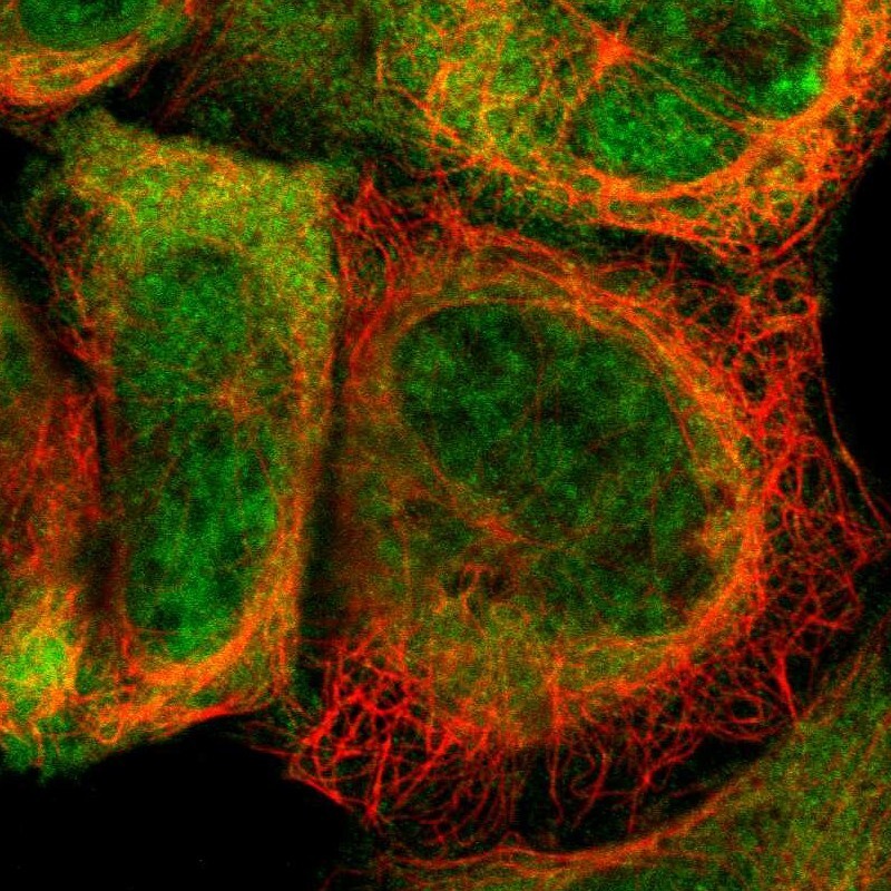
- Immunofluorescent staining of human cell line A-431 shows localization to nucleoplasm & cytosol.
- Sample type
- Human
Supportive validation
- Submitted by
- Atlas Antibodies (provider)
- Main image
- Experimental details
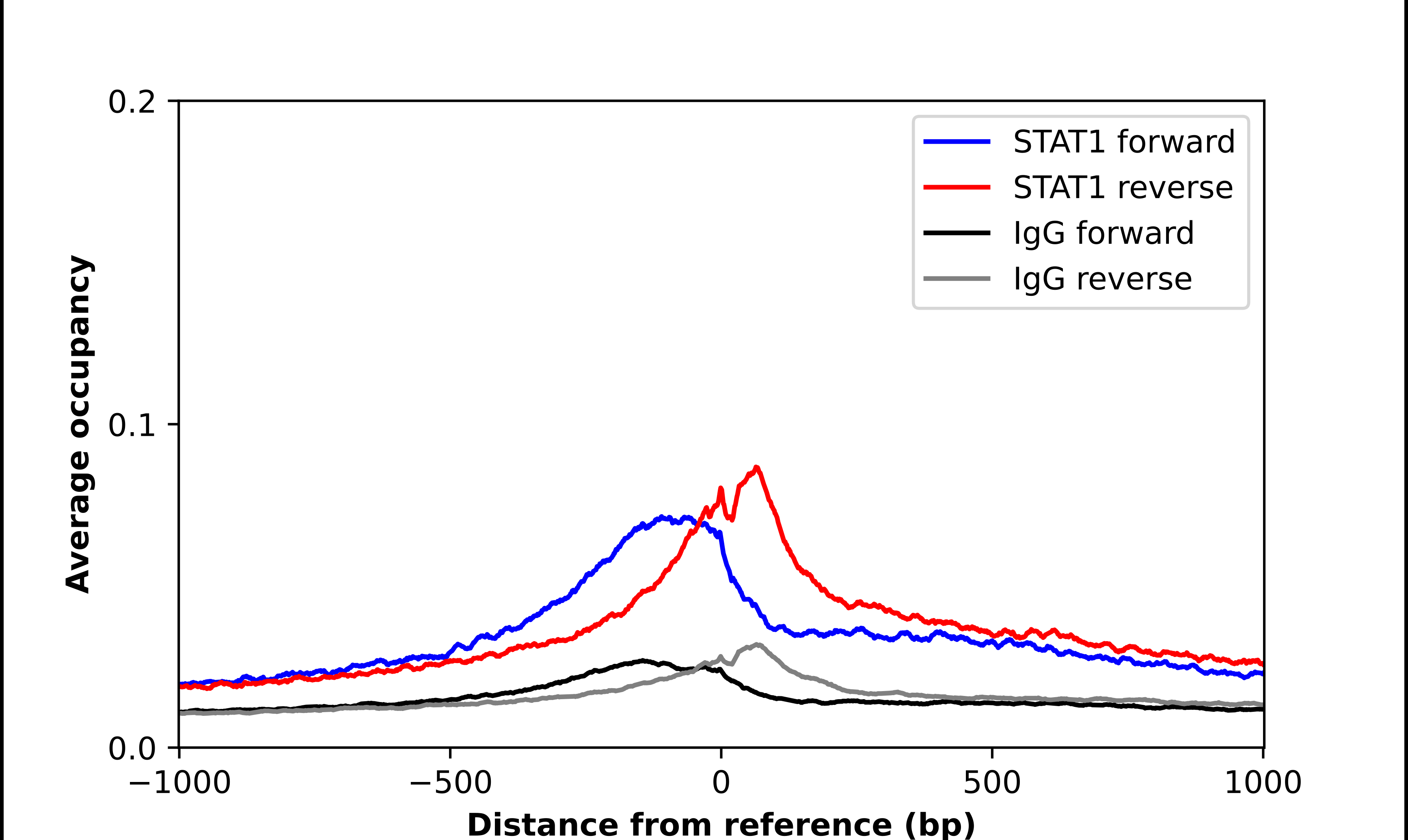
- ChIP-Exo-Seq composite graph for Anti-STAT1 (HPA000982, Lot 000053776) tested in K562 cells. Strand-specific reads (blue: forward, red: reverse) and IgG controls (black: forward, grey: reverse) are plotted against the distance from a composite set of reference binding sites. The antibody exhibits robust target enrichment compared to a non-specific IgG control and precisely reveals its structural organization around the binding site. Data generated by Prof. B. F. Pugh´s Lab at Cornell University.