Explore
 Validate
 Learn
Learn Western blot
 Immunoprecipitation
Antibody data
- Antibody Data
- Antigen structure
- References [3]
- Comments [0]
- Validations
- Western blot [2]
- Immunocytochemistry [3]
- Other assay [2]
Submit
Validation data
Reference
Comment
Report error
- Product number
- MA1-037X - Provider product page

- Provider
- Invitrogen Antibodies
- Product name
- STAT1 Monoclonal Antibody (15H3)
- Antibody type
- Monoclonal
- Antigen
- Other
- Description
- Note: This antibody requires overnight incubation in a Western blot for optimal performance. Do not use this antibody in a Fast Western blotting procedure.
- Reactivity
- Human
- Host
- Mouse
- Isotype
- IgG
- Antibody clone number
- 15H3
- Vial size
- 20 µL
- Concentration
- 1 mg/mL
- Storage
- -20°C
Submitted references A multi-shRNA vector enhances the silencing efficiency of exogenous and endogenous genes in human cells.
Dipeptidyl Peptidase-4 and Adolescent Idiopathic Scoliosis: Expression in Osteoblasts.
An Autocrine Cytokine/JAK/STAT-Signaling Induces Kynurenine Synthesis in Multidrug Resistant Human Cancer Cells.
Weng Y, Shi Y, Xia X, Zhou W, Wang H, Wang C
Oncology letters 2017 Mar;13(3):1553-1562
Oncology letters 2017 Mar;13(3):1553-1562
Dipeptidyl Peptidase-4 and Adolescent Idiopathic Scoliosis: Expression in Osteoblasts.
Normand E, Franco A, Moreau A, Marcil V
Scientific reports 2017 Jun 9;7(1):3173
Scientific reports 2017 Jun 9;7(1):3173
An Autocrine Cytokine/JAK/STAT-Signaling Induces Kynurenine Synthesis in Multidrug Resistant Human Cancer Cells.
Campia I, Buondonno I, Castella B, Rolando B, Kopecka J, Gazzano E, Ghigo D, Riganti C
PloS one 2015;10(5):e0126159
PloS one 2015;10(5):e0126159
No comments: Submit comment
Supportive validation
- Submitted by
- Invitrogen Antibodies (provider)
- Main image
- Experimental details
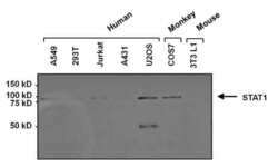
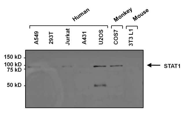
- Western blot analysis of STAT1 was performed by loading 25 µg of various whole cell lysates onto a 4-20% Tris-HCl polyacrylamide gel. Proteins were transferred to a PVDF membrane and blocked with 5% Milk/TBST for at least 1 hour. Membranes were probed with a mouse monoclonal antibody recognizing STAT1 (Product # MA1-037) at a dilution of 1:1000 overnight at 4°C on a rocking platform. Membranes were washed in TBS-0.1% Tween 20 and probed with a goat anti-mouse-HRP secondary antibody (Product # 31430) at a dilution of 1:20,000 for at least one hour. Membranes were washed and chemiluminescent detection performed using Super Signal West Dura (Product # 34075).

- Submitted by
- Invitrogen Antibodies (provider)
- Main image

- Experimental details
- Western blot analysis of STAT1 was performed by loading 25 µg of various whole cell lysates onto a 4-20% Tris-HCl polyacrylamide gel. Proteins were transferred to a PVDF membrane and blocked with 5% Milk/TBST for at least 1 hour. Membranes were probed with a mouse monoclonal antibody recognizing STAT1 (Product # MA1-037) at a dilution of 1:1000 overnight at 4°C on a rocking platform. Membranes were washed in TBS-0.1% Tween 20 and probed with a goat anti-mouse-HRP secondary antibody (Product # 31430) at a dilution of 1:20,000 for at least one hour. Membranes were washed and chemiluminescent detection performed using Super Signal West Dura (Product # 34075).
Supportive validation
- Submitted by
- Invitrogen Antibodies (provider)
- Main image

- Experimental details
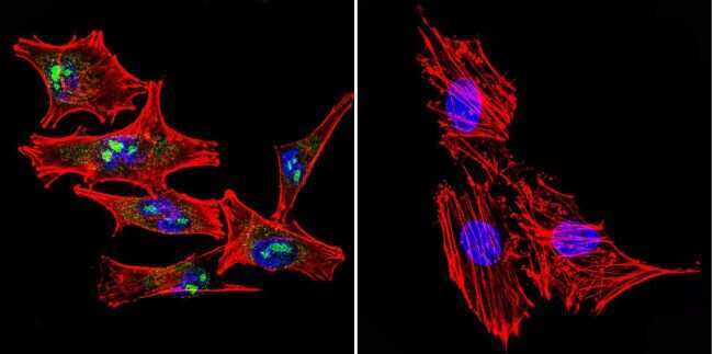
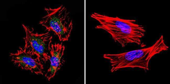
- Immunofluorescent analysis of STAT1 in A2058 Cells. Cells were grown on chamber slides and fixed with formaldehyde prior to staining. Cells were probed without (control) or with a STAT1 monoclonal antibody (Product # MA1-037) at a dilution of 1:20 overnight at 4 C, washed with PBS and incubated with a DyLight-488 conjugated secondary antibody (Product # 35503). STAT1 staining (green), F-Actin staining with Phalloidin (red) and nuclei with DAPI (blue) is shown. Images were taken at 60X magnification.
- Submitted by
- Invitrogen Antibodies (provider)
- Main image

- Experimental details
- Immunofluorescent analysis of STAT1 in HeLa Cells. Cells were grown on chamber slides and fixed with formaldehyde prior to staining. Cells were probed without (control) or with a STAT1 monoclonal antibody (Product # MA1-037) at a dilution of 1:20 overnight at 4 C, washed with PBS and incubated with a DyLight-488 conjugated secondary antibody (Product # 35503). STAT1 staining (green), F-Actin staining with Phalloidin (red) and nuclei with DAPI (blue) is shown. Images were taken at 60X magnification.
- Submitted by
- Invitrogen Antibodies (provider)
- Main image

- Experimental details
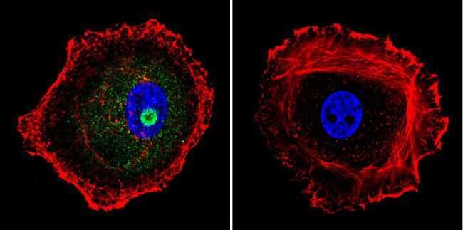
- Immunofluorescent analysis of STAT1 in MCF-7 Cells. Cells were grown on chamber slides and fixed with formaldehyde prior to staining. Cells were probed without (control) or with a STAT1 monoclonal antibody (Product # MA1-037) at a dilution of 1:20 overnight at 4 C, washed with PBS and incubated with a DyLight-488 conjugated secondary antibody (Product # 35503). STAT1 staining (green), F-Actin staining with Phalloidin (red) and nuclei with DAPI (blue) is shown. Images were taken at 60X magnification.
Supportive validation
- Submitted by
- Invitrogen Antibodies (provider)
- Main image

- Experimental details
- Immunoprecipitation of STAT1 was performed on U2OS cells. The antigen:antibody complex was formed by incubating 750 µg whole cell lysate with 2 µg of mouse monoclonal antibody recognizing STAT1 (Product # MA1-037) overnight on a rocking platform at 4°C. The immune-complex was then captured on 50 µl Protein A/G Plus Agarose (Product # 20423). Captured immune-complexes were then washed extensively and proteins eluted with 5X Reducing Sample Loading Dye (Product # 39000). Samples were then resolved on a 4-20% Tris-HCl polyacrylamide gel. Proteins were transferred to PVDF membrane and blocked with 5% Milk/TBS-0.1%Tween for at least 1 hour. Membranes were then probed with a mouse monoclonal antibody recognizing STAT1 (Product # MA1-037) at a dilution of 1:1000 overnight rotating at 4°C. Membranes were washed in TBS-0.1% Tween 20 and probed with a goat anti-mouse-HRP secondary antibody (Product # 31430) at a dilution of 1:20,000 for at least one hour. Membranes were washed and chemiluminescent detection performed using Super Signal West Dura (Product # 34075).

- Submitted by
- Invitrogen Antibodies (provider)
- Main image

- Experimental details
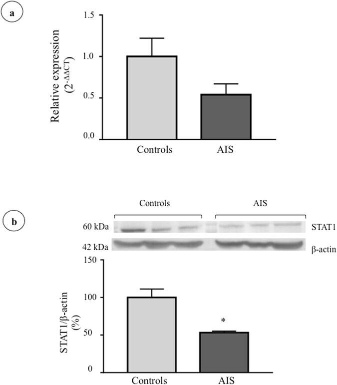
- Figure 6 STAT1 gene and protein expression in osteoblasts isolated from control and AIS patients. ( a ) STAT1 gene expression was measured in osteoblasts of controls (n = 7) and AIS patients (n = 13) by RT-qPCR with GAPDH as endogenous control. Relative expression was analyzed with the 2 -DeltaDeltaCT method. ( b ) STAT-1 protein expression was evaluated by Western blot (n = 3/group) and beta-actin was used as endogenous control. *P < 0.05 using two-tailed Student's t-test. Data are presented as mean +- standard deviation.