 Explore
 Validate
 Learn
Learn Western blot
 Chromatin Immunoprecipitation
Antibody data
- Antibody Data
- Antigen structure
- References [0]
- Comments [0]
- Validations
- Western blot [4]
- Immunocytochemistry [2]
- Flow cytometry [1]
Submit
Validation data
Reference
Comment
Report error
- Product number
- PA5-17635 - Provider product page

- Provider
- Invitrogen Antibodies
- Product name
- Phospho-STAT1 (Ser727) Polyclonal Antibody
- Antibody type
- Polyclonal
- Antigen
- Synthetic peptide
- Description
- It is not recommended to aliquot this antibody.
- Reactivity
- Human, Mouse, Rat
- Host
- Rabbit
- Isotype
- IgG
- Vial size
- 100 µL
- Concentration
- 56.5 µg/mL
- Storage
- -20°C
No comments: Submit comment
Supportive validation
- Submitted by
- Invitrogen Antibodies (provider)
- Main image

- Experimental details
- Western blot analysis of Phospho-STAT1 (pSer727) was performed by loading 30 µg of whole cell lysates from THP-1 cells either left untreated (UT, left lane) or stimulated with 1mM H2O2 (right lane) for 15 min, per well onto an SDS-PAGE gel. Proteins were transferred to a PVDF membrane and blocked with 5% non-fat dry milk in TBST for 1 hour at room temperature. The membrane was probed with a Phospho-STAT1 (pSer727) polyclonal antibody (Product # PA5-17635) at a dilution of 1:500 overnight at 4°C, washed in TBST, and probed with an HRP-conjugated goat anti-rabbit IgG secondary antibody at a dilution of 1:40,000 for 1 hour at room temperature. Detection was performed using ECL substrate. Data courtesy of the Innovators Program.

- Submitted by
- Invitrogen Antibodies (provider)
- Main image

- Experimental details
- Western blot analysis was performed on whole cell extracts (30 µg lysate) of MDA-MB-231 (Lane 1), MDA-MB-231 treated with IFN gamma (100ng/ml for 12 hr) (Lane 2), MCF7 (Lane 3) and MCF7 treated with IFN gamma (100ng/ml for 12 hr) (Lane 4). The blot was probed with Anti-Phospho-STAT1 (Ser727) Polyclonal Antibody (Product # PA5-17635, 1:1000 dilution) and detected by chemiluminescence using Goat anti-Rabbit IgG (H+L) Superclonal™ Secondary Antibody, HRP conjugate (Product # A27036, 0.25 µg/ml, 1:4000 dilution). A 91 kDa band corresponding to Phospho-STAT1 (Ser727) was observed across the cell lines tested and was enhanced upon treatment.

- Submitted by
- Invitrogen Antibodies (provider)
- Main image

- Experimental details
- Western blot analysis of Phospho-STAT1 (pSer727) was performed by loading 30 µg of whole cell lysates from THP-1 cells either left untreated (UT, left lane) or stimulated with 1mM H2O2 (right lane) for 15 min, per well onto an SDS-PAGE gel. Proteins were transferred to a PVDF membrane and blocked with 5% non-fat dry milk in TBST for 1 hour at room temperature. The membrane was probed with a Phospho-STAT1 (pSer727) polyclonal antibody (Product # PA5-17635) at a dilution of 1:500 overnight at 4°C, washed in TBST, and probed with an HRP-conjugated goat anti-rabbit IgG secondary antibody at a dilution of 1:40,000 for 1 hour at room temperature. Detection was performed using ECL substrate. Data courtesy of the Innovators Program.

- Submitted by
- Invitrogen Antibodies (provider)
- Main image

- Experimental details
- Western blot analysis was performed by loading 30 ug of HeLa (Lane 1), HeLa treated with IFN-gamma (1ng/ml for 15 minutes) (Lane 2), HeLa Cas9 (lane 3), HeLa Cas9 with IFN-gamma (1ng/ml for 15 minutes) (lane 4), HeLa JAK1 KO (Lane 5), HeLa JAK1 KO treated with (1ng/ml for 15 minutes)(Lane 6) whole cell extracts. The blots were probed with Phospho-STAT1 (Ser727) Polyclonal Antibody (Product: PA5-17635, 1:1000 dilution) and Goat anti-Rabbit IgG (H+L) Superclonal™ Secondary Antibody HRP conjugate (Product # A27036, 1:4000 dilution). Densitometric analysis of this western blot is shown in histogram (Fig. b). Reduction in signal in CRISPR mediated knockout (KO) confirms that antibody is specific to Phospho-STAT1

Supportive validation
- Submitted by
- Invitrogen Antibodies (provider)
- Main image
- Experimental details
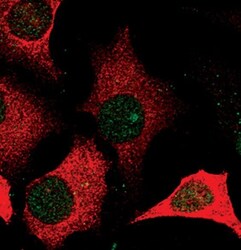
- Immunofluorescent analysis of Phospho-Stat1 pSer727 (green) in untreated HeLa cells. Cells fixed for 15 minutes in 4% formaldehyde were permeabilized with methanol and blocked with 5% normal goat serum + 0.3% Triton X-100 for 1 hour. Cells were probed with a Phospho-STAT1 pSer727 polyclonal antibody (Product # PA5-17635) at a dilution of 1:100 overnight at 4°C, washed with PBS, and incubated with a fluorescent-conjugated anti-rabbit IgG secondary antibody for at least 1 hour. Cells were also stained with an antibody against keratin (red).

- Submitted by
- Invitrogen Antibodies (provider)
- Main image
- Experimental details
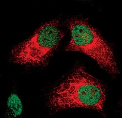
- Immunofluorescent analysis of Phospho-Stat1 pSer727 (green) in IFN-gamma-treated HeLa cells. Cells fixed for 15 minutes in 4% formaldehyde were permeabilized with methanol and blocked with 5% normal goat serum + 0.3% Triton X-100 for 1 hour. Cells were probed with a Phospho-STAT1 pSer727 polyclonal antibody (Product # PA5-17635) at a dilution of 1:100 overnight at 4°C, washed with PBS, and incubated with a fluorescently-conjugated anti-rabbit IgG secondary antibody for at least 1 hour. Cells were also stained with an antibody against keratin (red).

Supportive validation
- Submitted by
- Invitrogen Antibodies (provider)
- Main image

- Experimental details
- Flow cytometric analysis of Phospho-STAT1 pSer727 on HeLa cells left untreated (blue histogram), UV-treated (green histogram), or treated with phosphatase (red histogram). Cells were harvested, fixed with 4% formaldehyde for 10 minutes at 37°C, permeabilized in 90% ice-cold methanol for 30 minutes on ice, and incubated with a Phospho-STAT1 pSer727 polyclonal antibody (Product # PA5-17635) at 1:100 dilution 1 hour. For flow analysis, a 30-minute incubation with a fluorescently-conjugated secondary antibody was performed and cell staining was analyzed on a flow cytometer.
