Explore
 Validate
 Learn
Learn Western blot
Antibody data
- Antibody Data
- Antigen structure
- References [2]
- Comments [0]
- Validations
- Western blot [3]
- Immunoprecipitation [1]
Submit
Validation data
Reference
Comment
Report error
- Product number
- PAF-ST1 - Provider product page

- Provider
- R&D Systems
- Product name
- Human/Mouse STAT1 p91 Antibody
- Antibody type
- Polyclonal
- Description
- Antigen Affinity-purified. Detects human and mouse STAT1 p91 in Western blots. In Western blots, less than 1% cross-reactivity with human or mouse STAT1 p84 is observed. Additionally, no cross-reactivity with the other STATs has been detected by immunoprecipitation or Western blotting.
- Reactivity
- Human, Mouse
- Host
- Goat
- Conjugate
- Unconjugated
- Isotype
- IgG
- Vial size
- 100 ug
- Concentration
- LYOPH
- Storage
- Use a manual defrost freezer and avoid repeated freeze-thaw cycles. 12 months from date of receipt, -20 to -70 °C as supplied. 1 month, 2 to 8 °C under sterile conditions after reconstitution. 6 months, -20 to -70 °C under sterile conditions after reconstitution.
Submitted references Loss of ADAR1 in tumours overcomes resistance to immune checkpoint blockade.
Mediator Kinase Phosphorylation of STAT1 S727 Promotes Growth of Neoplasms With JAK-STAT Activation.
Ishizuka JJ, Manguso RT, Cheruiyot CK, Bi K, Panda A, Iracheta-Vellve A, Miller BC, Du PP, Yates KB, Dubrot J, Buchumenski I, Comstock DE, Brown FD, Ayer A, Kohnle IC, Pope HW, Zimmer MD, Sen DR, Lane-Reticker SK, Robitschek EJ, Griffin GK, Collins NB, Long AH, Doench JG, Kozono D, Levanon EY, Haining WN
Nature 2019 Jan;565(7737):43-48
Nature 2019 Jan;565(7737):43-48
Mediator Kinase Phosphorylation of STAT1 S727 Promotes Growth of Neoplasms With JAK-STAT Activation.
Nitulescu II, Meyer SC, Wen QJ, Crispino JD, Lemieux ME, Levine RL, Pelish HE, Shair MD
EBioMedicine 2017 Dec;26:112-125
EBioMedicine 2017 Dec;26:112-125
No comments: Submit comment
Supportive validation
- Submitted by
- R&D Systems (provider)
- Main image
- Experimental details
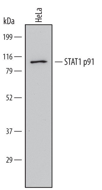
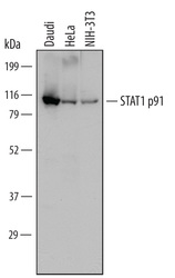
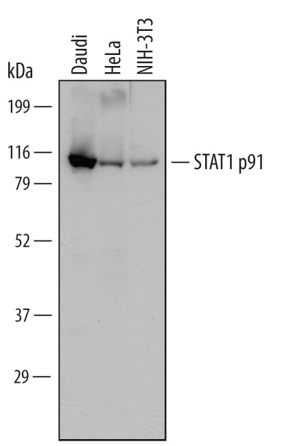
- Detection of Human and Mouse STAT1 p91 by Western Blot. Western blot shows lysates of Daudi human Burkitt's lymphoma cell line, HeLa human cervical epithelial carcinoma cell line, and NIH-3T3 mouse embryonic fibroblast cell line. PVDF Membrane was probed with 1 µg/mL of Goat Anti-Human/Mouse STAT1 p91 Antigen Affinity-purified Polyclonal Antibody (Catalog # PAF-ST1) followed by HRP-conjugated Anti-Goat IgG Secondary Antibody (Catalog # HAF109). A specific band was detected for STAT1 p91 at approximately 91 kDa (as indicated). This experiment was conducted under reducing conditions and using Immunoblot Buffer Group 1.
- Submitted by
- R&D Systems (provider)
- Main image
- Experimental details
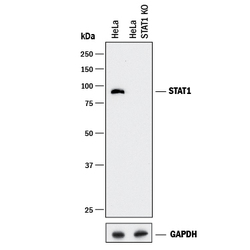
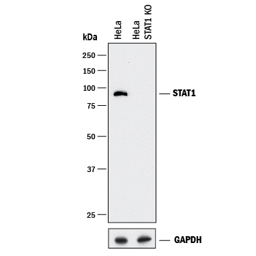
- Western Blot Shows Human STAT1 Specificity by Using Knockout Cell Line. Western blot shows lysates of HeLa human cervical epithelial carcinoma parental cell line and STAT1 knockout HeLa cell line (KO). PVDF membrane was probed with 0.25 µg/mL of Goat Anti-Human/Mouse STAT1 p91 Antigen Affinity-purified Polyclonal Antibody (Catalog # PAF-ST1) followed by HRP-conjugated Anti-Goat IgG Secondary Antibody (Catalog # HAF017). A specific band was detected for STAT1 at approximately 90 kDa (as indicated) in the parental HeLa cell line, but is not detectable in knockout HeLa cell line. GAPDH (Catalog # AF5718) is shown as a loading control. This experiment was conducted under reducing conditions and using Immunoblot Buffer Group 1.
- Submitted by
- R&D Systems (provider)
- Main image
- Experimental details
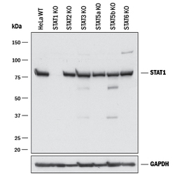
- Western Blot Shows Human STAT1 Specificity by Using Knockout Cell Line. Western blot shows lysates of HeLa human cervical epithelial carcinoma parental cell line, STAT1 knockout (KO) HeLa cell line, STAT2 KO HeLa cell line, STAT3 KO HeLa cell line, STAT5a KO HeLa cell line, STAT5b KO HeLa cell line, and STAT6 KO HeLa cell line. PVDF membrane was probed with 0.25 µg/mL of Goat Anti-Human/Mouse STAT1 p91 Antigen Affinity-purified Polyclonal Antibody (Catalog # PAF-ST1) followed by HRP-conjugated Anti-Goat IgG Secondary Antibody (Catalog # HAF017). A specific band was detected for STAT1 at approximately 90 kDa (as indicated) in the parental HeLa cell line, but is not detectable in knockout HeLa cell line. GAPDH (Catalog # AF5718) is shown as a loading control. This experiment was conducted under reducing conditions and using Immunoblot Buffer Group 1.
Supportive validation
- Submitted by
- R&D Systems (provider)
- Main image

- Experimental details
- Immunoprecipitation of Human STAT1. STAT1 p91 was immunoprecipitated from 200 μg of cell lysate of HeLa human cervical epithelial carcinoma cell line following incubation with 3 µg Goat Anti-Human/Mouse STAT1 p91 Antigen Affinity-purified Polyclonal Antibody (Catalog # PAF-ST1) overnight at 4 °C. STAT1 p91-antibody complexes were absorbed using Protein G sepharose (Invitrogen, Catalog # 10-1242). Immunoprecipitated STAT1 p91 was detected by Western blot using 1 µg/mL Human STAT1 Monoclonal Antibody (Catalog # MAB14901). View our recommended buffer recipes for immunoprecipitation.