Explore
 Validate
 Learn
Learn Western blot
 Immunocytochemistry
Antibody data
- Antibody Data
- Antigen structure
- References [1]
- Comments [0]
- Validations
- Immunocytochemistry [2]
- Immunoprecipitation [1]
- Immunohistochemistry [2]
- Other assay [2]
Submit
Validation data
Reference
Comment
Report error
- Product number
- PA5-85549 - Provider product page

- Provider
- Invitrogen Antibodies
- Product name
- DNMT3B Polyclonal Antibody
- Antibody type
- Polyclonal
- Antigen
- Recombinant full-length protein
- Description
- Keep as concentrated solution. Predicted reactivity: Pig (85%). Positive Control: HeLa, HeLa nuclear, GFP-tagged DNMT3B-transfested 293T. Store product as a concentrated solution. Centrifuge briefly prior to opening the vial.
- Reactivity
- Human, Rat
- Host
- Rabbit
- Isotype
- IgG
- Vial size
- 100 μL
- Concentration
- 1.29 mg/mL
- Storage
- Store at 4°C short term. For long term storage, store at -20°C, avoiding freeze/thaw cycles.
Submitted references PROSER1 mediates TET2 O-GlcNAcylation to regulate DNA demethylation on UTX-dependent enhancers and CpG islands.
Wang X, Rosikiewicz W, Sedkov Y, Martinez T, Hansen BS, Schreiner P, Christensen J, Xu B, Pruett-Miller SM, Helin K, Herz HM
Life science alliance 2022 Jan;5(1)
Life science alliance 2022 Jan;5(1)
No comments: Submit comment
Supportive validation
- Submitted by
- Invitrogen Antibodies (provider)
- Main image

- Experimental details
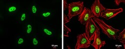
- DNMT3B Polyclonal Antibody detects DNMT3B protein at nucleus by immunofluorescent analysis. Sample: HeLa cells were fixed in 4% paraformaldehyde at RT for 15 min. Green: DNMT3B protein stained by DNMT3B Polyclonal Antibody (Product # PA5-85549) diluted at 1:200. Red: phalloidin, a cytoskeleton marker, diluted at 1:50. Scale bar = 10 µm.

- Submitted by
- Invitrogen Antibodies (provider)
- Main image
- Experimental details
- DNMT3B Polyclonal Antibody detects DNMT3B protein at nucleus by immunofluorescent analysis. Sample: HeLa cells were fixed in 4% paraformaldehyde at RT for 15 min. Green: DNMT3B protein stained by DNMT3B Polyclonal Antibody (Product # PA5-85549) diluted at 1:200. Red: phalloidin, a cytoskeleton marker, diluted at 1:50. Scale bar = 10 µm.

Supportive validation
- Submitted by
- Invitrogen Antibodies (provider)
- Main image

- Experimental details
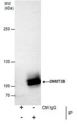
- Immunoprecipitation analysis of DNMT3B in K562 nuclear extracts with DNMT3B polyclonal antibody (Product # PA5-85549) using 5 µg of sample, followed by anti-Rabbit IgG secondary antibody.

Supportive validation
- Submitted by
- Invitrogen Antibodies (provider)
- Main image

- Experimental details
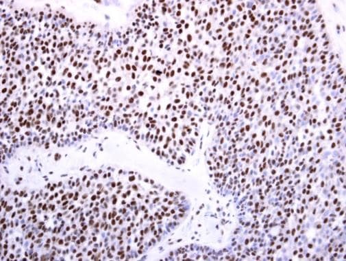
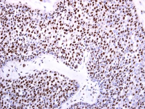
- Immunohistochemistry analysis of DNMT3B in paraffin-embedded human lung carcinoma using DNMT3B polyclonal antibody (Product # PA5-85549) at a dilution of 1:250.
- Submitted by
- Invitrogen Antibodies (provider)
- Main image

- Experimental details
- DNMT3B Polyclonal Antibody detects DNMT3B protein at nucleus on human lung carcinoma by immunohistochemical analysis. Sample: Paraffin-embedded human lung carcinoma. DNMT3B Polyclonal Antibody (Product # PA5-85549) diluted at 1:250. Antigen Retrieval: EDTA based buffer, pH 8.0, 15 min.
Supportive validation
- Submitted by
- Invitrogen Antibodies (provider)
- Main image
- Experimental details
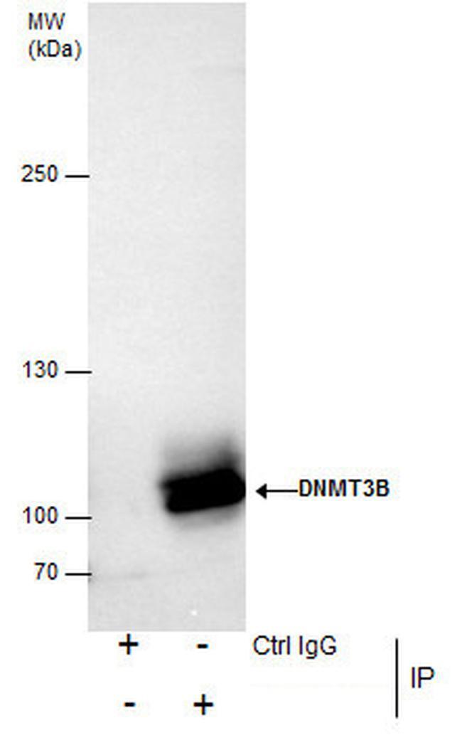
- Immunoprecipitation analysis of DNMT3B in K562 nuclear extracts with DNMT3B polyclonal antibody (Product # PA5-85549) using 5 µg of sample, followed by anti-Rabbit IgG secondary antibody.
- Submitted by
- Invitrogen Antibodies (provider)
- Main image
- Experimental details
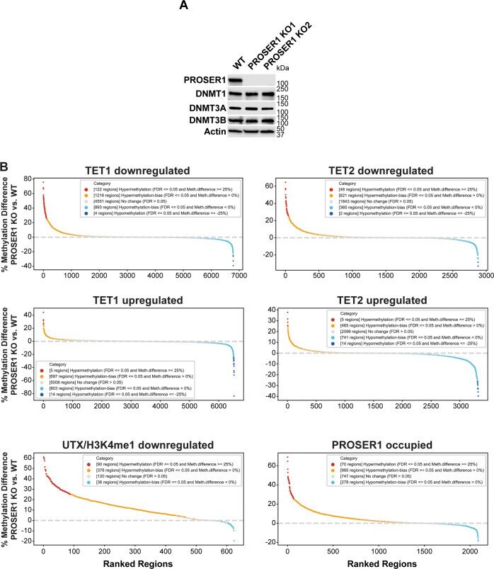
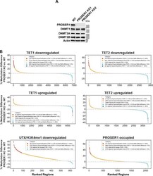
- Figure S7. Exploring alternative mechanisms of PROSER1-mediated changes in DNA methylation. (A) Western blot for members of the DNA methyltransferase family from nuclear extracts of WT and PROSER1 KO HEK293 cells. Actin was used as a loading control. (B) DNA methylation rank plots showing regions associated with TET1 down-regulation, TET2 down-regulation, TET1 up-regulation, TET2 up-regulation, UTX/H3K4me1 down-regulation, and PROSER1-occupied regions in PROSER1 KO versus WT HEK293 cells (all FC >= 2). Regions with CpGs covered by at least 10 whole genome bisulfite sequencing reads (please see ""whole genome bisulfite sequencing data processing"" in the ""Materials and Methods"" section for further details) were ranked from hyper- to hypomethylated on the x-axis.