 Explore
 Validate
 Learn
Learn Western blot
Antibody data
- Antibody Data
- Antigen structure
- References [10]
- Comments [0]
- Validations
- Western blot [1]
- Immunohistochemistry [3]
- Other assay [6]
Submit
Validation data
Reference
Comment
Report error
- Product number
- PA1-884 - Provider product page

- Provider
- Invitrogen Antibodies
- Product name
- DNMT3B Polyclonal Antibody
- Antibody type
- Polyclonal
- Antigen
- Synthetic peptide
- Description
- PA1-884 detects DNA methyltransferase 3 from human and mouse tissues and cells as well as recombinant human Dnmt3. This antibody has been used to detect both recombinant and endogenous DNMT3b and recombinant DNMT3a. PA1-884 has been successfully used in Immunohistochemistry (paraffin) and Western blot procedures. By Western blot, this antibody detects DNMT3 from P19 nuclear extracts. The PA1-884 immunogen is a synthetic peptide corresponding to residues 1-14 of mouse DNMT3b. This sequence is 79% conserved between human and mouse. PA1-884 immunizing peptide (Cat. # PEP-117) is available for use in neutralization and control experiments.
- Reactivity
- Human, Mouse
- Host
- Rabbit
- Isotype
- IgG
- Vial size
- 100 µg
- Concentration
- 1 mg/mL
- Storage
- -20° C, Avoid Freeze/Thaw Cycles
Submitted references Epigenetic Regulation of CDH1 Is Altered after HOXB7-Silencing in MDA-MB-468 Triple-Negative Breast Cancer Cells.
Dnmt3b catalytic activity is critical for its tumour suppressor function in lymphomagenesis and is associated with c-Met oncogenic signalling.
Decreases in different Dnmt3b activities drive distinct development of hematologic malignancies in mice.
Catalytically inactive Dnmt3b rescues mouse embryonic development by accessory and repressive functions.
Diabetes impairs wound healing by Dnmt1-dependent dysregulation of hematopoietic stem cells differentiation towards macrophages.
The SNF2 family ATPase LSH promotes cell-autonomous de novo DNA methylation in somatic cells.
G9a/GLP Complex Maintains Imprinted DNA Methylation in Embryonic Stem Cells.
LSH and G9a/GLP complex are required for developmentally programmed DNA methylation.
De novo DNA methyltransferase DNMT3b interacts with NEDD8-modified proteins.
LSH cooperates with DNA methyltransferases to repress transcription.
Paço A, Leitão-Castro J, Freitas R
Genes 2021 Oct 3;12(10)
Genes 2021 Oct 3;12(10)
Dnmt3b catalytic activity is critical for its tumour suppressor function in lymphomagenesis and is associated with c-Met oncogenic signalling.
Lopusna K, Nowialis P, Opavska J, Abraham A, Riva A, Opavsky R
EBioMedicine 2021 Jan;63:103191
EBioMedicine 2021 Jan;63:103191
Decreases in different Dnmt3b activities drive distinct development of hematologic malignancies in mice.
Lopusna K, Nowialis P, Opavska J, Abraham A, Riva A, Haney SL, Opavsky R
The Journal of biological chemistry 2021 Jan-Jun;296:100285
The Journal of biological chemistry 2021 Jan-Jun;296:100285
Catalytically inactive Dnmt3b rescues mouse embryonic development by accessory and repressive functions.
Nowialis P, Lopusna K, Opavska J, Haney SL, Abraham A, Sheng P, Riva A, Natarajan A, Guryanova O, Simpson M, Hlady R, Xie M, Opavsky R
Nature communications 2019 Sep 26;10(1):4374
Nature communications 2019 Sep 26;10(1):4374
Diabetes impairs wound healing by Dnmt1-dependent dysregulation of hematopoietic stem cells differentiation towards macrophages.
Yan J, Tie G, Wang S, Tutto A, DeMarco N, Khair L, Fazzio TG, Messina LM
Nature communications 2018 Jan 2;9(1):33
Nature communications 2018 Jan 2;9(1):33
The SNF2 family ATPase LSH promotes cell-autonomous de novo DNA methylation in somatic cells.
Termanis A, Torrea N, Culley J, Kerr A, Ramsahoye B, Stancheva I
Nucleic acids research 2016 Sep 19;44(16):7592-604
Nucleic acids research 2016 Sep 19;44(16):7592-604
G9a/GLP Complex Maintains Imprinted DNA Methylation in Embryonic Stem Cells.
Zhang T, Termanis A, Özkan B, Bao XX, Culley J, de Lima Alves F, Rappsilber J, Ramsahoye B, Stancheva I
Cell reports 2016 Apr 5;15(1):77-85
Cell reports 2016 Apr 5;15(1):77-85
LSH and G9a/GLP complex are required for developmentally programmed DNA methylation.
Myant K, Termanis A, Sundaram AY, Boe T, Li C, Merusi C, Burrage J, de Las Heras JI, Stancheva I
Genome research 2011 Jan;21(1):83-94
Genome research 2011 Jan;21(1):83-94
De novo DNA methyltransferase DNMT3b interacts with NEDD8-modified proteins.
Shamay M, Greenway M, Liao G, Ambinder RF, Hayward SD
The Journal of biological chemistry 2010 Nov 19;285(47):36377-86
The Journal of biological chemistry 2010 Nov 19;285(47):36377-86
LSH cooperates with DNA methyltransferases to repress transcription.
Myant K, Stancheva I
Molecular and cellular biology 2008 Jan;28(1):215-26
Molecular and cellular biology 2008 Jan;28(1):215-26
No comments: Submit comment
Supportive validation
- Submitted by
- Invitrogen Antibodies (provider)
- Main image

- Experimental details
- Western blot analysis of DNA Methyltransferase 3 was performed by loading 25 µg of Hela cell lysates and a molecular weight protein ladder onto an SDS polyacrylamide gel. Proteins were transferred to a PVDF membrane and blocked with a blocking buffer at 4ºC overnight. The membrane was probed with a DNA Methyltransferase 3 polyclonal antibody (Product # PA1-884) at a dilution of 1:1000 overnight at 4°C, washed in TBST, and probed with an HRP-conjugated secondary antibody for 1 hr at room temperature in the dark. Results show a band at 97 kDa.

Supportive validation
- Submitted by
- Invitrogen Antibodies (provider)
- Main image

- Experimental details
- Immunohistochemistry analysis of DNA Methyltransferase 3 showing positive staining in the nucleus and cytoplasm of paraffin-treated Human breast carcinoma (right) compared with a negative control in the absence of primary antibody (left). To expose target proteins, antigen retrieval method was performed using 10mM sodium citrate (pH 6.0) microwaved for 8-15 min. Following antigen retrieval, tissues were blocked in 3% H2O2-methanol for 15 min at room temperature, washed with ddH2O and PBS, and then probed with a DNA Methyltransferase 3 polyclonal antibody (Product # PA1-884) diluted by 3% BSA-PBS at a dilution of 1:500 overnight at 4°C in a humidified chamber. Tissues were washed extensively PBST and detection was performed using an HRP-conjugated secondary antibody followed by colorimetric detection using a DAB kit. Tissues were counterstained with hematoxylin and dehydrated with ethanol and xylene to prep for mounting.

- Submitted by
- Invitrogen Antibodies (provider)
- Main image

- Experimental details
- Immunohistochemistry analysis of DNA Methyltransferase 3 showing positive staining in the nucleus of paraffin-treated Human testis tissue (right) compared with a negative control in the absence of primary antibody (left). To expose target proteins, antigen retrieval method was performed using 10mM sodium citrate (pH 6.0) microwaved for 8-15 min. Following antigen retrieval, tissues were blocked in 3% H2O2-methanol for 15 min at room temperature, washed with ddH2O and PBS, and then probed with a DNA Methyltransferase 3 polyclonal antibody (Product # PA1-884) diluted by 3% BSA-PBS at a dilution of 1:500 overnight at 4°C in a humidified chamber. Tissues were washed extensively PBST and detection was performed using an HRP-conjugated secondary antibody followed by colorimetric detection using a DAB kit. Tissues were counterstained with hematoxylin and dehydrated with ethanol and xylene to prep for mounting.

- Submitted by
- Invitrogen Antibodies (provider)
- Main image

- Experimental details
- Immunohistochemistry analysis of DNA Methyltransferase 3 showing positive staining in the nucleus and cytoplasm of paraffin-treated Mouse brain tissue (right) compared with a negative control in the absence of primary antibody (left). To expose target proteins, antigen retrieval method was performed using 10mM sodium citrate (pH 6.0) microwaved for 8-15 min. Following antigen retrieval, tissues were blocked in 3% H2O2-methanol for 15 min at room temperature, washed with ddH2O and PBS, and then probed with a DNA Methyltransferase 3 polyclonal antibody (Product # PA1-884) diluted by 3% BSA-PBS at a dilution of 1:200 overnight at 4°C in a humidified chamber. Tissues were washed extensively PBST and detection was performed using an HRP-conjugated secondary antibody followed by colorimetric detection using a DAB kit. Tissues were counterstained with hematoxylin and dehydrated with ethanol and xylene to prep for mounting.

Supportive validation
- Submitted by
- Invitrogen Antibodies (provider)
- Main image
- Experimental details
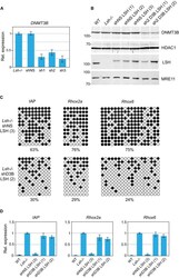
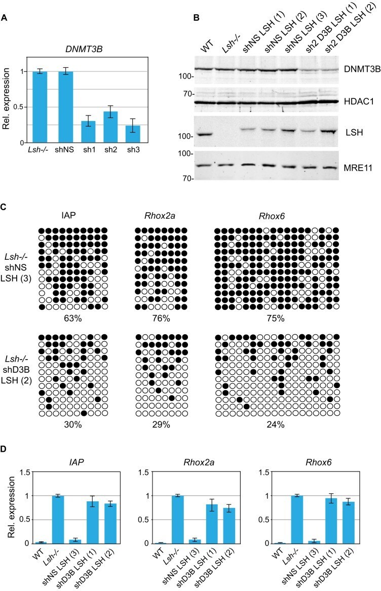
- Figure 6. DNMT3B is required for de novo DNA methylation of IAPs and gene promoters upon LSH expression in Lsh-/- MEFs. ( A ) Expression of DNMT3B in Lsh-/- MEFs expressing either a non-silencing shRNA (shNS) or shRNAs (sh1, sh2 and sh3) targeting DNMT3B as assessed by quantitative RT-PCR. The error bars denote standard deviation. ( B ) Western blots showing the presence of 3xFLAG-tagged wild-type LSH in Lsh-/- clonal MEF lines expressing either non-silencing shRNA (shNS) or shRNA targeting DNMT3B (sh2 D3B). HDAC1 and MRE11 were used as loading controls. ( C ) Bisulfite DNA sequencing of IAPs and the promoters of Rhox2a and Rhox6 genes in indicated cell lines. Methylated CpGs are shown in black. ( D ) Expression relative to Gapdh of IAP s, Rhox2a and Rhox6 in indicated cell lines. The expression detected in Lsh-/- MEFs was designated as 1. The error bars denote standard deviation.
- Submitted by
- Invitrogen Antibodies (provider)
- Main image
- Experimental details
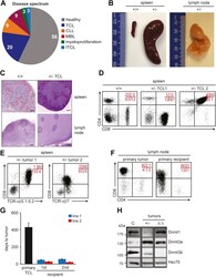
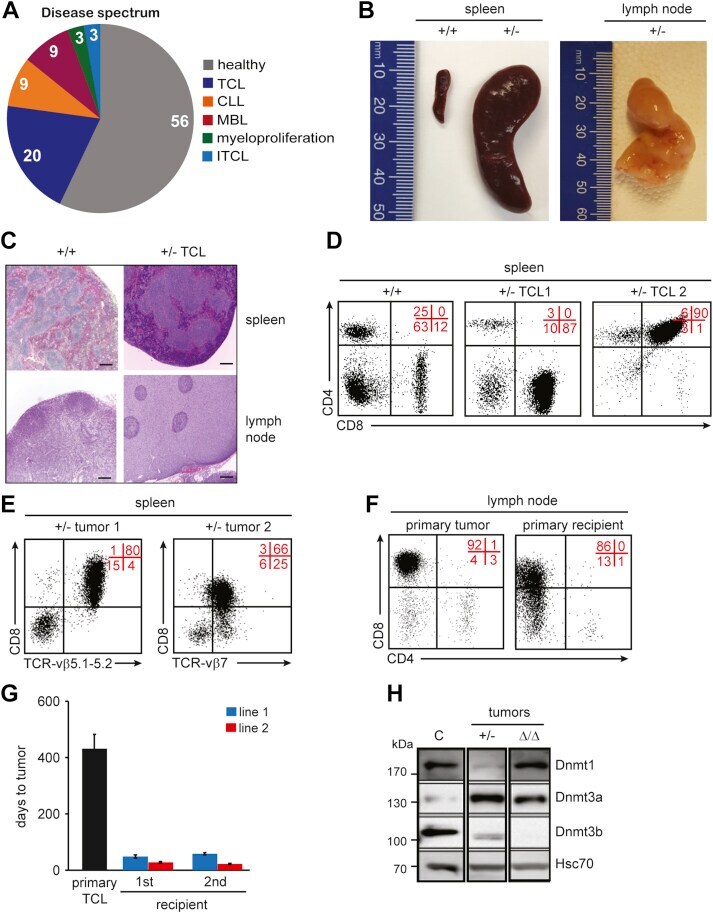
- Figure 1 Majority of Dnmt3b +/- mice develop T-cell lymphomas. A , Disease spectrum observed in Dnmt3b +/- mice (n = 35) determined by FACS. Values present percentage of mice diagnosed with indicated disease. B , Representative image of healthy spleen of Dnmt3b +/+ (+/+) mice and spleen and lymph node of terminally ill Dnmt3b +/- (+/-) mice that developed TCL. C , Histological staining of spleen and lymph node of age-matched Dnmt3b +/+ control ( +/+ ) and terminally ill Dnmt3b +/- mouse (+/- TCL) (magnification 40x). D , Representative FACS diagrams of CD4 and CD8 expression in cells isolated from the spleen of healthy Dnmt3b +/+ mice (+/+) and terminally sick Dnmt3b +/- mice that developed CD8+ (+/- TCL1) and CD4+CD8+ (+/- TCL2) lymphomas. Quadrant statistics are indicated in red here and in all figures. E , Representative FACS diagrams showing clonal TCR-vbeta expression in Dnmt3b +/- lymphomas (+/- tumor 1 and 2). F , Representative FACS diagrams of CD4 and CD8 expression in cells isolated from tumors that developed in terminally sick Dnmt3b +/- mice (primary tumor) and sublethally irradiated FVB-recipient mice injected with primary lymphoma (primary recipient). G , Time to tumor development for primary mice (primary TCL), primary (first) and secondary (second) sublethally irradiated FVB-recipient mice serially transplanted with primary TCL isolated from the lymph nodes of terminally sick Dnmt3b +/- mice. Data are presented as average time to tumor development. Two TCL lin
- Submitted by
- Invitrogen Antibodies (provider)
- Main image

- Experimental details
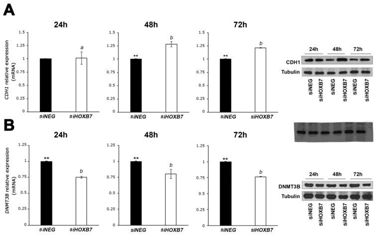
- Figure 3 Expression analyses of CDH1 ( A ) and DNMT3B ( B ) in MDA-MB-468 cells transfected with siHOXB7 and siNEG analyzed by RT-qPCR and western blots at three time points post-transfection. Three independent biological replicates were used in the statistical analyses comparing two variants (siNEG and siHOXB7) and the significant differences indicated with asterisks based on the analyses using t -test. The differences in the expression measures at the three time points were compared using the post hoc Tukey test. CDH1 expression was significantly higher in siHOXB7-transfected cells than in controls at 48 h and 72 h (**, p < 0.05). Comparing the three time points, the expression of CDH1 is significantly different at 24 h in these cells ( a ) in comparison with 48 h and 72 h ( b ). No significant differences in CDH1 expression were detected between 48 h and 72 h. DNMT3B expression was significantly lower in HOXB7-silenced cells than in the controls in the three time points analyzed, and no significant differences were detected in the expression between these time points ( b ). In the western-blot analyses, beta-tubulin was used as the reference protein. a : significant expression differences between time-points in the siHOXB7 cells evaluated with post hoc Tukey test; b : no significant expression differences between time-points in the siHOXB7 cells evaluated with post hoc Tukey test.
- Submitted by
- Invitrogen Antibodies (provider)
- Main image
- Experimental details
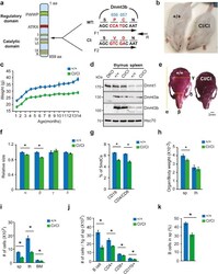
- Fig. 1 Mice expressing catalytically inactive Dnmt3b develop ICF-like phenotype. a. (left) Schematic representation of mouse Dnmt3b protein structure. Regulatory domain containing PWWP and ATRX domains and catalytic domain with methyltransferase motifs (I, IV, VI, IX and X) are depicted (right) . Nucleotide and amino acid sequences of exon 19 in wild-type (WT) and Dnmt3b CI (CI) allele. Amino acid substitution (P656V and C657D) in CI allele results in inactivation of the CA . Position of genotyping primers is indicated by arrows. F1-R detects wild-type sequence; F2-R detects mutated sequence. b. Micrograph of 6-week-old Dnmt3b +/+ (+/+) and Dnmt3b CI/CI (CI/CI) mice. c. Body weight values of Dnmt3b +/+ and Dnmt3b CI/CI mice at indicated ages ( n = 10). p

- Submitted by
- Invitrogen Antibodies (provider)
- Main image
- Experimental details
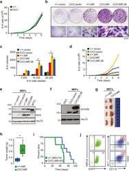
- Fig. 2 Loss of Dnmt3b's CA promotes cellular transformation in vitro and in vivo. a. Proliferation rate of Dnmt3b +/+ (+/+) and Dnmt3b CI/CI (CI/CI) MEFs cells. Representative results of one out of three experiments are shown. b. Colony formation assay of Dnmt3b +/+ and Dnmt3b CI/CI MEFs cells transduced with lentiviral control vector (+/+;vector and CI/CI;vector, respectively), MEFs transformed with vectors expressing c-Myc and Ras (+/+;MR and CI/CI;MR) and transformed Dnmt3b CI/CI MEFs transduced with lentiviral vector encoding Dnmt3b WT (CI/CI;MR;3B). Top pictures depicts culture plates with macroscopically visible colonies after fixation and crystal violet staining. Lower pictures show the same colonies at the microscopic level (8x magnification). Scale bar represents 2 mm. c. Quantification of number of colonies obtained from experiment described in (b). Cells were seeded at depicted numbers. Data are presented as mean +-SEM from three independent experiments. * p

- Submitted by
- Invitrogen Antibodies (provider)
- Main image

- Experimental details
- Fig. 3 Loss of Dnmt3b's CA accelerates MYC induced lymphomagenesis. a. KM survival curve of MYC;Dnmt3b +/+ (+/+, n = 20), MYC;Dnmt3b +/CI (+/CI, n = 13) and MYC;Dnmt3b CI/CI (CI/CI, n = 12) mice. p =3, p
