 Explore
 Validate
 Learn
Learn ELISA
ELISA Flow cytometry
Antibody data
- Antibody Data
- Antigen structure
- References [3]
- Comments [0]
- Validations
- Flow cytometry [1]
- Other assay [4]
Submit
Validation data
Reference
Comment
Report error
- Product number
- MA1-70054 - Provider product page

- Provider
- Invitrogen Antibodies
- Product name
- Complement C3b Monoclonal Antibody (10C7)
- Antibody type
- Monoclonal
- Antigen
- Other
- Description
- MA1-70054 detects C3/C3b/iC3b in human and mouse samples. MA1-70054 has been successfully used in flow cytometry and ELISA procedures. The MA1-70054 immunogen is C57BL/6 thymocytes incubated with a rat IgG2b anti-mouse Thy-1 (RmT1) and C57BL/6 serum (as a source for C3).
- Reactivity
- Human, Mouse
- Host
- Mouse
- Isotype
- IgG
- Antibody clone number
- 10C7
- Vial size
- 250 μg
- Concentration
- 1 mg/mL
- Storage
- Store at 4°C short term. For long term storage, store at -20°C, avoiding freeze/thaw cycles.
Submitted references Bacillus anthracis Poly-γ-D-Glutamate Capsule Inhibits Opsonic Phagocytosis by Impeding Complement Activation.
Human mesenchymal stromal cells undergo apoptosis and fragmentation after intravenous application in immune-competent mice.
Host iron status and erythropoietic response to iron supplementation determines susceptibility to the RBC stage of falciparum malaria during pregnancy.
Sharma S, Bhatnagar R, Gaur D
Frontiers in immunology 2020;11:462
Frontiers in immunology 2020;11:462
Human mesenchymal stromal cells undergo apoptosis and fragmentation after intravenous application in immune-competent mice.
Leibacher J, Dauber K, Ehser S, Brixner V, Kollar K, Vogel A, Spohn G, Schäfer R, Seifried E, Henschler R
Cytotherapy 2017 Jan;19(1):61-74
Cytotherapy 2017 Jan;19(1):61-74
Host iron status and erythropoietic response to iron supplementation determines susceptibility to the RBC stage of falciparum malaria during pregnancy.
Goheen MM, Bah A, Wegmüller R, Verhoef H, Darboe B, Danso E, Prentice AM, Cerami C
Scientific reports 2017 Dec 15;7(1):17674
Scientific reports 2017 Dec 15;7(1):17674
No comments: Submit comment
Supportive validation
- Submitted by
- Invitrogen Antibodies (provider)
- Main image
- Experimental details
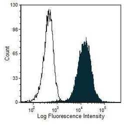
- Flow cytometric analysis of C57BL/6 mouse thymocytes incubated with rabbit anti-mouse T cells and then incubated with fresh human serum were stained using a Complement C3b monoclonal antibody (Product # MA1-70054) (filled histogram) or mouse IgG1 isotype control (open histogram).

Supportive validation
- Submitted by
- Invitrogen Antibodies (provider)
- Main image
- Experimental details
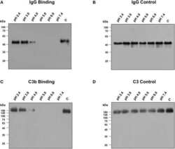
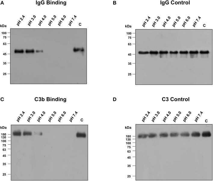
- Figure 6 Binding of IgG and C3b with encapsulated strain of Bacillus anthracis incubated at different pH buffers ranging from 2.4 to 7.4, supplemented with 10% normal human serum or 50 mug/ml of purified human IgG. (A) Immunoblot assay for the detection of purified IgG binding on encapsulated strain of B. anthracis . IgG binding was observed at pH closer to the isoelectric point of poly-gamma-D-glutamate (pI 3.2). (B) Purified IgG was stable at different pH buffers as detected by immunoblot. (C) Immunoblot assay for the detection of human C3b binding on encapsulated strain of B. anthracis . C3b binding was also observed at pH closer to the isoelectric point of poly-gamma-D-glutamate. (D) Normal human serum (NHS) was incubated at different pH buffers and C3 was observed to be stable at low pH as analyzed by immunoblot.
- Submitted by
- Invitrogen Antibodies (provider)
- Main image
- Experimental details
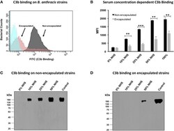
- Figure 2 C3b deposition on encapsulated and non-encapsulated strains of Bacillus anthracis . (A) Flow cytometry histogram of C3b deposition on encapsulated and non-encapsulated B. anthracis strains incubated in 10% human serum. Gray and pink shading indicates non-encapsulated and encapsulated bacteria, respectively. Blue shading indicates bacteria incubated in phosphate-buffered saline (PBS) alone. (B) Mean fluorescence index (MFI) of serum concentration-dependent C3b deposition on encapsulated and non-encapsulated bacteria. Black and gray bars represent non-encapsulated and encapsulated strains, respectively. The scatter plots for the corresponding histograms are represented in Figure S3 . (C,D) Immunoblot assays to detect serum concentration-dependent C3b deposition on non-encapsulated (C) and encapsulated strains (D) ; 1% normal human serum was used as positive control. Each data point represents the mean of three independent experiments. Error bars represent standard error of the mean. Statistical significance is highlighted by the following denotations: ** for P -value < 0.01, and *** for P -value < 0.001.

- Submitted by
- Invitrogen Antibodies (provider)
- Main image

- Experimental details
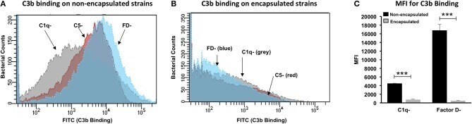
- Figure 4 Contributory role of classical and alternative pathways in C3b deposition on Bacillus anthracis . Flow cytometry histogram for C3b deposition on non-encapsulated (A) and encapsulated (B) B. anthracis strains incubated in 10% human serum deficient in complement component C1q (C1q-), factor D (FD-), and C5 (C5-). Gray, red, and blue histogram shading represents C3b deposition in C1q-, C5-, and FD- deficient sera, respectively. (C) C3b binding represented as mean fluorescence index (MFI) observed with non-encapsulated (black bars) and encapsulated (gray bars) B. anthracis strains incubated in C1q and FD sera. Each bar represents the mean of three independent experiments. Error bars represent standard error of the mean. Statistical significance is highlighted by the following denotations: *** for P -value < 0.001.
- Submitted by
- Invitrogen Antibodies (provider)
- Main image

- Experimental details
- Figure 6 Binding of IgG and C3b with encapsulated strain of Bacillus anthracis incubated at different pH buffers ranging from 2.4 to 7.4, supplemented with 10% normal human serum or 50 mug/ml of purified human IgG. (A) Immunoblot assay for the detection of purified IgG binding on encapsulated strain of B. anthracis . IgG binding was observed at pH closer to the isoelectric point of poly-gamma-D-glutamate (pI 3.2). (B) Purified IgG was stable at different pH buffers as detected by immunoblot. (C) Immunoblot assay for the detection of human C3b binding on encapsulated strain of B. anthracis . C3b binding was also observed at pH closer to the isoelectric point of poly-gamma-D-glutamate. (D) Normal human serum (NHS) was incubated at different pH buffers and C3 was observed to be stable at low pH as analyzed by immunoblot.
