Explore
 Validate
 Learn
Learn Western blot
 Other assay
Antibody data
- Antibody Data
- Antigen structure
- References [5]
- Comments [0]
- Validations
- Other assay [4]
Submit
Validation data
Reference
Comment
Report error
- Product number
- PA1-29715 - Provider product page

- Provider
- Invitrogen Antibodies
- Product name
- Complement C3 Polyclonal Antibody
- Antibody type
- Polyclonal
- Antigen
- Other
- Description
- Specificity: Monospecific to human C3 complement component. This antibody has also been reported to recognize the C3 fragments, C3a, C3b and C3c. Product is supplied as diluted unpurified antiserum. Store as a concentrated solution. Centrifuge briefly prior to opening the vial.
- Reactivity
- Human, Mouse, Rat, Guinea Pig, Hamster, Porcine, Rabbit
- Host
- Goat
- Isotype
- IgG
- Vial size
- 1 mL
- Concentration
- 89 mg/mL
- Storage
- Store at 4°C short term. For long term storage, store at -20°C, avoiding freeze/thaw cycles.
Submitted references Microglial- and Astrocyte-Specific Expression of Purinergic Signaling Components and Inflammatory Mediators in the Rat Hippocampus During Trimethyltin-Induced Neurodegeneration.
Podocytes Produce and Secrete Functional Complement C3 and Complement Factor H.
Prolonged intraocular residence and retinal tissue distribution of a fourth-generation compstatin-based C3 inhibitor in non-human primates.
Induction of NTPDase1/CD39 by Reactive Microglia and Macrophages Is Associated With the Functional State During EAE.
The immunosuppression mechanism of hypodermin A on complement.
Dragić M, Mitrović N, Adžić M, Nedeljković N, Grković I
ASN neuro 2021 Jan-Dec;13:17590914211044882
ASN neuro 2021 Jan-Dec;13:17590914211044882
Podocytes Produce and Secrete Functional Complement C3 and Complement Factor H.
Mühlig AK, Keir LS, Abt JC, Heidelbach HS, Horton R, Welsh GI, Meyer-Schwesinger C, Licht C, Coward RJ, Fester L, Saleem MA, Oh J
Frontiers in immunology 2020;11:1833
Frontiers in immunology 2020;11:1833
Prolonged intraocular residence and retinal tissue distribution of a fourth-generation compstatin-based C3 inhibitor in non-human primates.
Hughes S, Gumas J, Lee R, Rumano M, Berger N, Gautam AK, Sfyroera G, Chan AL, Gnanaguru G, Connor KM, Kim BJ, Dunaief JL, Ricklin D, Hajishengallis G, Yancopoulou D, Reis ES, Mastellos DC, Lambris JD
Clinical immunology (Orlando, Fla.) 2020 May;214:108391
Clinical immunology (Orlando, Fla.) 2020 May;214:108391
Induction of NTPDase1/CD39 by Reactive Microglia and Macrophages Is Associated With the Functional State During EAE.
Jakovljevic M, Lavrnja I, Bozic I, Milosevic A, Bjelobaba I, Savic D, Sévigny J, Pekovic S, Nedeljkovic N, Laketa D
Frontiers in neuroscience 2019;13:410
Frontiers in neuroscience 2019;13:410
The immunosuppression mechanism of hypodermin A on complement.
Chen RJ, Hu AK, Yuan HH, Wang ZZ, Ji DJ, Wu LL, Zhang TY, Zhu YH, Sun W, Zhu XR
Parasitology international 2014 Apr;63(2):392-6
Parasitology international 2014 Apr;63(2):392-6
No comments: Submit comment
Supportive validation
- Submitted by
- Invitrogen Antibodies (provider)
- Main image
- Experimental details
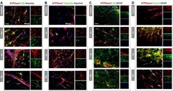
- FIGURE 8 Heterogeneity of NTPDase1 positive astrocytes. Representative micrographs showing double immunofluorescence labeling directed to NTPDase1 ( red fluorescence ) and (A) glutamine synthetase - GS (B) vimentin, (C) Complement component 3 - C3, and (D) Cox-2 ( green fluorescence for all). Sections (A,B) were counterstained with Hoechst for cell nuclei ( blue fluorescence ). Sections (C,D) were co-labeled with GFAP ( blue fluorescence ). Scale bar applicable to all micrographs = 20 mum.

- Submitted by
- Invitrogen Antibodies (provider)
- Main image

- Experimental details
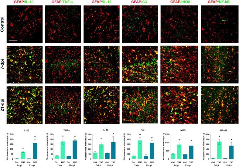
- Figure 7. Assessment of the functional state of reactive astrocytes after TMT exposure. Double immunofluorescent staining of GFAP and IL-1beta, TNF-alpha, IL-10, C3, iNOS and NF-kB and corresponding integrated fluorescence density expressed as AUs +- SEM in the injured CA area at 7 and 21 dpi. Significance shown inside the graphs: * p < .05 or less compared to age-match Ctrl. Note . AU = arbitrary unit; Ctrl = control; dpi = days post intoxication; GFAP = glial fibrillary acidic protein; IL-10 = interleukin-10; IL-1beta = interleukin-1beta; iNOS = inducible nitric oxide synthase; NF-kB = nuclear factor-kB; SEM = standard error of the mean; TMT = trimethyltin; TNF-alpha = tumor necrosis factor alpha.
- Submitted by
- Invitrogen Antibodies (provider)
- Main image

- Experimental details
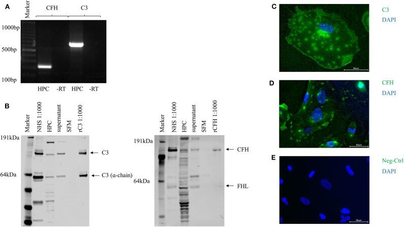
- Figure 1 Complement proteins C3 and CFH were expressed and secreted by unstimulated human podocytes (HPC). (A) Detection of PCR products for C3 (320 bp) and CFH (783 bp) in conventional reverse transcriptase PCR compared to -RT control (-RT, control of cDNA without addition of RT; bp, basepairs). (B) Protein expression for C3 and CFH was determined by Western blot from whole cell podocyte lysate (HPC) and cell culture supernatant after 24 h in serum-free media (SFM). Normal human serum (NHS, 1:1,000) and recombinant CFH or C3 were used as positive controls (rC3/rCFH, 1:1,000), SFM was used as negative control. In the C3 Western blot also the alpha-chain of C3 can be detected. The CFH gene has two protein products: CFH and factor H like-protein (FHL). Both are detected in this Western blot in the podocyte cell lysate, in NHS and to a small extend in the cell culture supernatant (representative Western blots, n = 4). (C) Protein expression of C3 and (D) CFH was confirmed in immunofluorescence on the surface of non-permeabilized cultured podocytes compared to isotype negative control (Neg-Ctrl) (E) . ( n = 3, C3 and CFH = green, nucleus = blue, scale bar 50 mum, 40x).
- Submitted by
- Invitrogen Antibodies (provider)
- Main image

- Experimental details
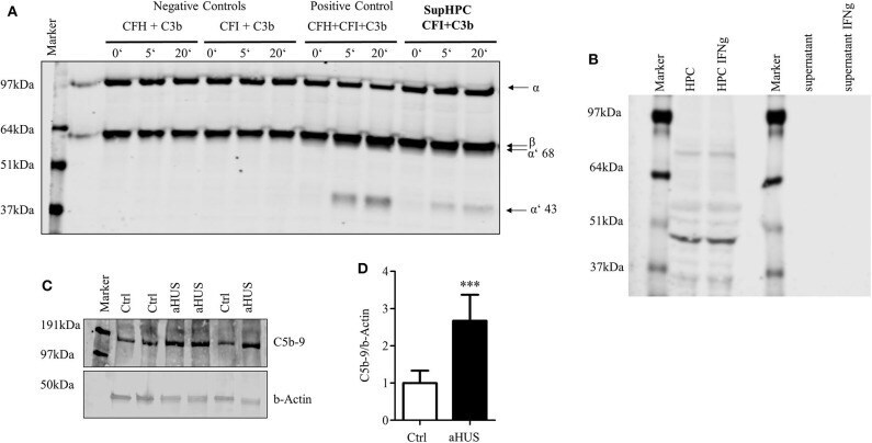
- Figure 4 Podocyte-secreted CFH functionally active. Functionality of secreted CFH was tested in a cofactor assay and in a C5b-9 deposition assay in podocytes with lost CFH derived complement control. (A) Cofactor assay: CFH together with CFI can split recombinant C3b. Split products with a size of 68 and 43 kDa can be detected in Western blot against C3. Podocyte supernatant with added CFI was able to cleave added recombinant C3b. Positive control: C3b, CFI and CFH were added to serum free media (SFM) in supersaturated amounts, leading to immediate cleavage of C3b (illustrated by the appearance of the cleavage products of alpha'68- and alpha'43 kD). There is no detection of any cleavage products after the incubation of C3b and CFI, or C3b and CFI in SFM (left part). When C3b and CFI are added to podocyte supernatant, C3b split products can be detected. (B) Podocyte lysate (HPC) shows a clear signal for CD46 as a membrane bound complement regulator, but did not shed CD46 into the supernatant. The treatment of podocytes with interferon gamma (IFNg, 10 ng/mL, 24 h) did not result in an induction of CD46 in the cell lysate or enhanced shedding in the cell culture supernatant (representative Western blot of 3 independent experiments). (C,D) The deposition of terminal complement complex C5b-9 was compared on normal human podocytes and podocytes from a patient suffering from atypical hemolytic uremic syndrome (aHUS). Patient's CFH was not able to bind to the cell surface. Complement