Explore
 Validate
 Learn
Learn Western blot
Antibody data
- Antibody Data
- Antigen structure
- References [7]
- Comments [0]
- Validations
- Western blot [2]
Submit
Validation data
Reference
Comment
Report error
- Product number
- AF1650 - Provider product page

- Provider
- Novus Biologicals
- Product name
- Rabbit Polyclonal Caspase-8 Antibody
- Antibody type
- Polyclonal
- Description
- Antigen Affinity-purified. Detects human/mouse Caspase-8 and cleavage products. Detects multiple isoforms of Caspase-8.
- Reactivity
- Human, Mouse
- Host
- Rabbit
- Conjugate
- Unconjugated
- Isotype
- IgG
- Vial size
- 100 ug
- Concentration
- LYOPH
- Storage
- Use a manual defrost freezer and avoid repeated freeze-thaw cycles. 12 months from date of receipt, -20 to -70 degreesC as supplied. 1 month, 2 to 8 degreesC under sterile conditions after reconstitution. 6 months, -20 to -70 degreesC under sterile conditions after reconstitution.
Submitted references Ferrichrome identified from Lactobacillus casei ATCC334 induces apoptosis through its iron-binding site in gastric cancer cells.
The expression of the Sprouty 1 protein inversely correlates with growth, proliferation, migration and invasion of ovarian cancer cells.
NRBF2 regulates autophagy and prevents liver injury by modulating Atg14L-linked phosphatidylinositol-3 kinase III activity.
TCEA3 binds to TGF-beta receptor I and induces Smad-independent, JNK-dependent apoptosis in ovarian cancer cells.
Hyperactivation of the mammalian degenerin MDEG promotes caspase-8 activation and apoptosis.
Inhibition of protein degradation induces apoptosis through a microtubule-associated protein 1 light chain 3-mediated activation of caspase-8 at intracellular membranes.
Theiler's virus-induced intrinsic apoptosis in M1-D macrophages is Bax mediated and restricts virus infectivity: a mechanism for persistence of a cytolytic virus.
Ijiri M, Fujiya M, Konishi H, Tanaka H, Ueno N, Kashima S, Moriichi K, Sasajima J, Ikuta K, Okumura T
Tumour biology : the journal of the International Society for Oncodevelopmental Biology and Medicine 2017 Jun;39(6):1010428317711311
Tumour biology : the journal of the International Society for Oncodevelopmental Biology and Medicine 2017 Jun;39(6):1010428317711311
The expression of the Sprouty 1 protein inversely correlates with growth, proliferation, migration and invasion of ovarian cancer cells.
Masoumi-Moghaddam S, Amini A, Ehteda A, Wei AQ, Morris DL
Journal of ovarian research 2014;7:61
Journal of ovarian research 2014;7:61
NRBF2 regulates autophagy and prevents liver injury by modulating Atg14L-linked phosphatidylinositol-3 kinase III activity.
Lu J, He L, Behrends C, Araki M, Araki K, Jun Wang Q, Catanzaro JM, Friedman SL, Zong WX, Fiel MI, Li M, Yue Z
Nature communications 2014 May 22;5:3920
Nature communications 2014 May 22;5:3920
TCEA3 binds to TGF-beta receptor I and induces Smad-independent, JNK-dependent apoptosis in ovarian cancer cells.
Cha Y, Kim DK, Hyun J, Kim SJ, Park KS
Cellular signalling 2013 May;25(5):1245-51
Cellular signalling 2013 May;25(5):1245-51
Hyperactivation of the mammalian degenerin MDEG promotes caspase-8 activation and apoptosis.
Pan JA, Fan Y, Gandhirajan RK, Madesh M, Zong WX
The Journal of biological chemistry 2013 Feb 1;288(5):2952-63
The Journal of biological chemistry 2013 Feb 1;288(5):2952-63
Inhibition of protein degradation induces apoptosis through a microtubule-associated protein 1 light chain 3-mediated activation of caspase-8 at intracellular membranes.
Pan JA, Ullman E, Dou Z, Zong WX
Molecular and cellular biology 2011 Aug;31(15):3158-70
Molecular and cellular biology 2011 Aug;31(15):3158-70
Theiler's virus-induced intrinsic apoptosis in M1-D macrophages is Bax mediated and restricts virus infectivity: a mechanism for persistence of a cytolytic virus.
Son KN, Becker RP, Kallio P, Lipton HL
Journal of virology 2008 May;82(9):4502-10
Journal of virology 2008 May;82(9):4502-10
No comments: Submit comment
Supportive validation
- Submitted by
- Novus Biologicals (provider)
- Main image

- Experimental details
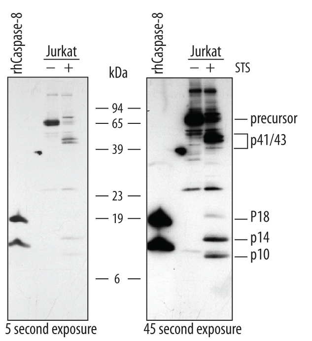
- Detection of Human Caspase-8 by Western Blot. Western blot shows lysates of Jurkat human acute T cell leukemia cell line untreated (-) or treated (+) with 1 mM staurosporine (STS) for for 3 hours. PVDF membrane was probed with 0.5 µg/mL of Rabbit Anti-Human/Mouse Caspase-8 Antigen Affinity-purified Polyclonal Antibody (Catalog # AF1650), followed by HRP-conjugated Anti-Rabbit IgG Secondary Antibody (Catalog # HAF008). For additional reference Recombinant Human Caspase-8 (Catalog # 705-C8) was included. Specific bands were detected for Caspase-8 precursor at approximately 57-60 kDa (as indicated). In STS-treated samples, specific bands were detected for Caspase-8 p41/43 subunit at approximately 41 and 43 kDa (as indicated) and Caspase-8 p18, p14, and p10 subunits at approximately 18 kDa, 14 kDa, and 10 kDa, respectively (as indicated). This experiment was conducted under reducing conditions and using Immunoblot Buffer Group 4.
- Submitted by
- Novus Biologicals (provider)
- Main image

- Experimental details
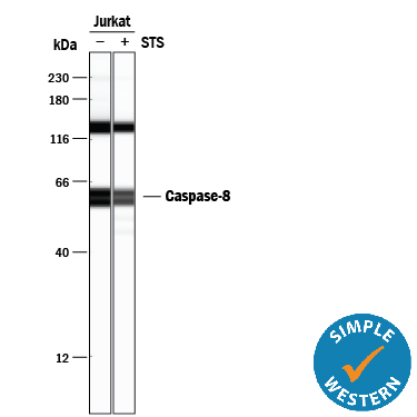
- Detection of Human Caspase-8 by Simple WesternTM. Simple Western lane view shows lysates of Jurkat human acute T cell leukemia cell line untreated (-) or treated (+) with 1 mM Staurosporine (STS) for 3 hours, loaded at 0.2 mg/mL. Specific bands were detected for Caspase-8 at approximately 58-62 kDa (as indicated) using 5 µg/mL of Rabbit Anti-Human/Mouse Caspase-8 Antigen Affinity-purified Polyclonal Antibody (Catalog # AF1650). This experiment was conducted under reducing conditions and using the 12-230 kDa separation system.