Explore
 Validate
 Learn
Learn Western blot
Antibody data
- Antibody Data
- Antigen structure
- References [4]
- Comments [0]
- Validations
- Western blot [1]
Submit
Validation data
Reference
Comment
Report error
- Product number
- MAB704 - Provider product page

- Provider
- Novus Biologicals
- Product name
- Mouse Monoclonal Caspase-8 Antibody
- Antibody type
- Monoclonal
- Description
- Protein A or G purified from ascites. Detects human Caspase-8 precursor in Western blots and a 42 kDa doublet generated during apoptosis.
- Reactivity
- Human
- Host
- Mouse
- Isotype
- IgG
- Vial size
- 100 ug
- Concentration
- LYOPH
- Storage
- Use a manual defrost freezer and avoid repeated freeze-thaw cycles. 12 months from date of receipt, -20 to -70 degreesC as supplied. 1 month, 2 to 8 degreesC under sterile conditions after reconstitution. 6 months, -20 to -70 degreesC under sterile conditions after reconstitution.
Submitted references Silencing of heat shock protein 27 increases the radiosensitivity of non‑small cell lung carcinoma cells.
Regulation of the extrinsic apoptotic pathway by the extracellular matrix glycoprotein EMILIN2.
Enhanced susceptibility to tumor necrosis factor-related apoptosis-inducing ligand-mediated apoptosis in oral squamous cell carcinoma cells treated with phosphatidylinositol 3-kinase inhibitors.
Changes in lamina structure are followed by spatial reorganization of heterochromatic regions in caspase-8-activated human mesenchymal stem cells.
Xu L, Lin X, Zheng Y, Zhou H
Molecular medicine reports 2019 Jul;20(1):613-621
Molecular medicine reports 2019 Jul;20(1):613-621
Regulation of the extrinsic apoptotic pathway by the extracellular matrix glycoprotein EMILIN2.
Mongiat M, Ligresti G, Marastoni S, Lorenzon E, Doliana R, Colombatti A
Molecular and cellular biology 2007 Oct;27(20):7176-87
Molecular and cellular biology 2007 Oct;27(20):7176-87
Enhanced susceptibility to tumor necrosis factor-related apoptosis-inducing ligand-mediated apoptosis in oral squamous cell carcinoma cells treated with phosphatidylinositol 3-kinase inhibitors.
Uchida M, Iwase M, Takaoka S, Yoshiba S, Kondo G, Watanabe H, Ohashi M, Nagumo M, Shintani S
International journal of oncology 2007 May;30(5):1163-71
International journal of oncology 2007 May;30(5):1163-71
Changes in lamina structure are followed by spatial reorganization of heterochromatic regions in caspase-8-activated human mesenchymal stem cells.
Raz V, Carlotti F, Vermolen BJ, van der Poel E, Sloos WC, Knaän-Shanzer S, de Vries AA, Hoeben RC, Young IT, Tanke HJ, Garini Y, Dirks RW
Journal of cell science 2006 Oct 15;119(Pt 20):4247-56
Journal of cell science 2006 Oct 15;119(Pt 20):4247-56
No comments: Submit comment
Supportive validation
- Submitted by
- Novus Biologicals (provider)
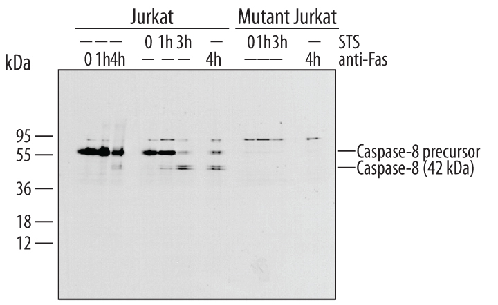
- Main image

- Experimental details
- Detection of Human Caspase-8 by Western Blot. Western blot shows lysates of Jurkat human acute T cell leukemia cell line untreated (-) or treated (+) with Human FAS Antigen Affinity-purified Polyclonal Antibody (Catalog # AF126) for indicated times. PVDF membrane was probed with 1 µg/mL of Human Caspase-8 Monoclonal Antibody (Catalog # MAB704), followed by HRP-conjugated Anti-Mouse IgG Secondary Antibody (Catalog # HAF007). Specific bands were detected for Caspase-8 at approximately 55 and 42 kDa (as indicated). This experiment was conducted under reducing conditions and using Immunoblot Buffer Group 4.