Explore
 Validate
 Learn
Learn Western blot
Antibody data
- Antibody Data
- Antigen structure
- References [0]
- Comments [0]
- Validations
- Western blot [2]
- Immunohistochemistry [2]
- Flow cytometry [2]
Submit
Validation data
Reference
Comment
Report error
- Product number
- 10-1019 - Provider product page

- Provider
- ABEOMICS Inc.
- Product name
- Anti-Caspase-8 Antibody
- Antibody type
- Monoclonal
- Description
- Apoptosis occurs during normal cellular development and involves dramatic changes in cellular structure. Disruption of apoptosis may contribute to cancer as well as other autoimmune diseases. Caspase family of cysteine proteases has been shown to play a key role in apoptosis. Caspase-8 is a 55 kDa cytosolic protein that is synthesized as an inactive pro-enzyme. Activation of caspase-8 involves a two-step proteolysis: the cleavage of caspase-8 to generate a 43 and a 12 kDa fragment which is further processed to 10 kDa. The p43 is then cleaved to yield p26 and the release of the active site containing p18.The Active/Cleaved Caspase-8 polyclonal antisera recognizes the large and small subunits of active/cleaved caspase-8. Whereas the antisera has a strong preference for active/cleaved caspase-8, in some cell or tissue systems or techniques the antisera may also recognize the proform of caspase-8 as well as intermediate caspase-8 cleavage fragments.
- Reactivity
- Human, Mouse
- Host
- Mouse
- Conjugate
- Unconjugated
- Antigen sequence
A partial length recombinant protei
n  (a.a 179-385) of Caspase-8  was
used as the immunogen for this anti
body.- Isotype
- IgG
- Antibody clone number
- ABM14C1
- Vial size
- 100 µg
- Concentration
- 0.5 mg/ml
- Storage
- Store the antibody at 4°C, stable for 6 months. For long-term storage, store at -20°C. Avoid repeat freez thawing
No comments: Submit comment
Supportive validation
- Submitted by
- ABEOMICS Inc. (provider)
- Main image
- Experimental details
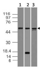
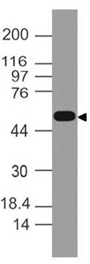
- Western blot analysis of Caspase-8. Anti-Caspase-8 antibody (Clone: ABM14C1) was used at 2 µg/ml on Molt-4, Kato III and HepG2 lysates.
- Protocol
- Protocol

- Submitted by
- ABEOMICS Inc. (provider)
- Main image
- Experimental details
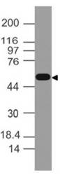
- Western blot analysis of Caspase-8. Anti-Caspase-8 antibody (Clone: ABM14C1) was used at 4 µg/ml on EL-4 lysate.
- Protocol
- Protocol
Supportive validation
- Submitted by
- ABEOMICS Inc. (provider)
- Main image
- Experimental details
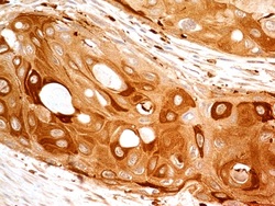
- Immunohistochemical analysis of Caspase-8 in human Esophagus using anti-Caspase-8 antibody (Clone: ABM14C1) at 5 µg/ml.
- Protocol
- Protocol

- Submitted by
- ABEOMICS Inc. (provider)
- Main image

- Experimental details
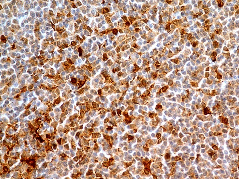
- Immunohistochemical analysis of Caspase-8 in human Tonsil using anti-Caspase-8 antibody (Clone: ABM14C1) at 5 µg/ml.
- Protocol
- Protocol
Supportive validation
- Submitted by
- ABEOMICS Inc. (provider)
- Main image
- Experimental details
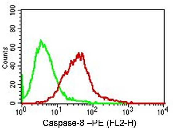
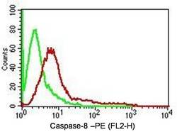
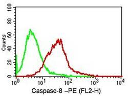
- Intracellular Flow analysis of Caspase-8 antibody in Hela cells using 0.5 µg/ 10^6 cells of anti-Caspase-8 antibody (ABM14C1). Green represents isotype control; red represents anti-Caspase-8 antibody. Goat anti-mouse PE conjugate was used as secondary antibody. (Cells were fixed with 4% paraformaldehyde for 10 min and washed with PBS by centrifuging at 1100 for 5 min followed by permeabilization for 20 min and washed again as mentioned above. Then cell were incubated with primary antibody for 45 min. and after washing the cells twice in PBS, incubated with conjugated secondary antibody for 30 min. Data acquisition was done after washing twice with PBS as mentioned above).
- Protocol
- Protocol
- Submitted by
- ABEOMICS Inc. (provider)
- Main image
- Experimental details
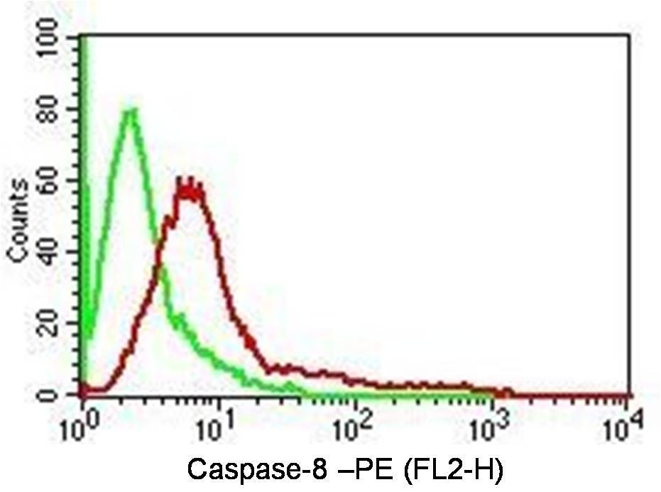
- Intracellular Flow analysis of Caspase-8 antibody in Jurkat cells using 0.5 µg/ 10^6 cells of anti-Caspase-8 antibody (ABM14C1). Green represents isotype control; red represents anti-Caspase-8 antibody. Goat anti-mouse PE conjugate was used as secondary antibody. (Cells were fixed with 4% paraformaldehyde for 10 min and washed with PBS by centrifuging at 1100 for 5 min followed by permeabilization for 20 min and washed again as mentioned above. Then cell were incubated with primary antibody for 45 min. and after washing the cells twice in PBS, incubated with conjugated secondary antibody for 30 min. Data acquisition was done after washing twice with PBS as mentioned above).
- Protocol
- Protocol