 Explore
 Validate
 Learn
Learn Western blot
Antibody data
- Antibody Data
- Antigen structure
- References [7]
- Comments [0]
- Validations
- Western blot [4]
- Immunohistochemistry [1]
- Flow cytometry [1]
Submit
Validation data
Reference
Comment
Report error
- Product number
- NB100-56527 - Provider product page

- Provider
- Novus Biologicals
- Proper citation
- Novus Cat#NB100-56527, RRID:AB_837869
- Product name
- Mouse Monoclonal Caspase-8 Antibody
- Antibody type
- Monoclonal
- Description
- Protein G purified. This can be used for detection of the pro-form of Caspase-8.
- Reactivity
- Human, Simian
- Host
- Mouse
- Isotype
- IgG
- Vial size
- 0.1 mg
- Concentration
- 1.0 mg/ml
- Storage
- Store at 4C short term. Aliquot and store at -20C long term. Avoid freeze-thaw cycles.
Submitted references Mutational landscape of penile squamous cell carcinoma in a Chinese population.
The induction of apoptosis and autophagy by Wasabia japonica extract in colon cancer.
Simultaneous Host-Pathogen Transcriptome Analysis during Granulibacter bethesdensis Infection of Neutrophils from Healthy Subjects and Patients with Chronic Granulomatous Disease.
Potent anti-cancer effect of 3'-hydroxypterostilbene in human colon xenograft tumors.
Inhibition of HSP27 alone or in combination with pAKT inhibition as therapeutic approaches to target SPARC-induced glioma cell survival.
Helenalin suppresses essential immune functions of activated CD4+ T cells by multiple mechanisms.
Proteasome inhibition activates the mitochondrial pathway of apoptosis in human CD4+ T cells.
Wang Y, Wang K, Chen Y, Zhou J, Liang Y, Yang X, Li X, Cao Y, Wang D, Luo L, Li B, Li D, Wang L, Liang Z, Gao C, Wang Q, Lv Q, Li Z, Shi Y, Niu H
International journal of cancer 2019 Sep 1;145(5):1280-1289
International journal of cancer 2019 Sep 1;145(5):1280-1289
The induction of apoptosis and autophagy by Wasabia japonica extract in colon cancer.
Hsuan SW, Chyau CC, Hung HY, Chen JH, Chou FP
European journal of nutrition 2016 Mar;55(2):491-503
European journal of nutrition 2016 Mar;55(2):491-503
Simultaneous Host-Pathogen Transcriptome Analysis during Granulibacter bethesdensis Infection of Neutrophils from Healthy Subjects and Patients with Chronic Granulomatous Disease.
Greenberg DE, Sturdevant DE, Marshall-Batty KR, Chu J, Pettinato AM, Virtaneva K, Lane J, Geller BL, Porcella SF, Gallin JI, Holland SM, Zarember KA
Infection and immunity 2015 Nov;83(11):4277-92
Infection and immunity 2015 Nov;83(11):4277-92
Potent anti-cancer effect of 3'-hydroxypterostilbene in human colon xenograft tumors.
Cheng TC, Lai CS, Chung MC, Kalyanam N, Majeed M, Ho CT, Ho YS, Pan MH
PloS one 2014;9(11):e111814
PloS one 2014;9(11):e111814
Inhibition of HSP27 alone or in combination with pAKT inhibition as therapeutic approaches to target SPARC-induced glioma cell survival.
Schultz CR, Golembieski WA, King DA, Brown SL, Brodie C, Rempel SA
Molecular cancer 2012 Apr 5;11:20
Molecular cancer 2012 Apr 5;11:20
Helenalin suppresses essential immune functions of activated CD4+ T cells by multiple mechanisms.
Berges C, Fuchs D, Opelz G, Daniel V, Naujokat C
Molecular immunology 2009 Sep;46(15):2892-901
Molecular immunology 2009 Sep;46(15):2892-901
Proteasome inhibition activates the mitochondrial pathway of apoptosis in human CD4+ T cells.
Berges C, Haberstock H, Fuchs D, Sadeghi M, Opelz G, Daniel V, Naujokat C
Journal of cellular biochemistry 2009 Nov 1;108(4):935-46
Journal of cellular biochemistry 2009 Nov 1;108(4):935-46
No comments: Submit comment
Supportive validation
- Submitted by
- Novus Biologicals (provider)
- Main image
- Experimental details
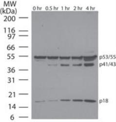
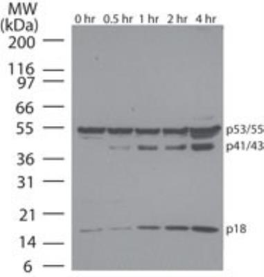
- Western Blot: Caspase-8 Antibody (90A992) [NB100-56527] - Western blot analysis of Caspase-8 in Jurkat cells using Caspase-8 antibody at 1 ug/ml. Cells were treated with 2 uM staurosporine for different time periods. Caspase-8 activation is detected in western blots by the presence of Caspase-8 cleavage fragments. The antibody detected both pro (full length) and active (cleaved) Caspase-8, depending on the treatment time points. A basal level of endogenously cleaved Caspase-8 can be see in untreated Jurkat cells. Goat anti-mouse lg HRP secondary antibody and PicoTect ECL substrate solution were used for this test.
- Submitted by
- Novus Biologicals (provider)
- Main image

- Experimental details
- Simple Western: Caspase-8 Antibody (90A992) [NB100-56527] - Simple Western lane view shows a specific band for Caspase 8 in 0.5 mg/ml of Hek293 lysate. This experiment was performed under reducing conditions using the 12-230 kDa separation system.

- Submitted by
- Novus Biologicals (provider)
- Main image
- Experimental details
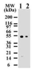
- Western Blot: Caspase-8 Antibody (90A992) [NB100-56527] - Analysis using the Biotin conjugate of NB100-56527. Detection of human Caspase-8 using Jurkat lysates with NB100-55786 at 2 ug/ml (lane 1) and 0.5 ug/ml (lane 2) dilution. NB100-55786 only detects 55 kDa Caspase-8 in Jurkat cells.

- Submitted by
- Novus Biologicals (provider)
- Main image
- Experimental details
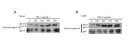
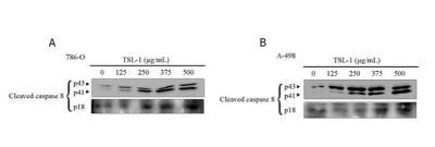
- Western Blot: Caspase-8 Antibody (90A992) [NB100-56527] - Caspase-8 expression in 786-O and A-498 cellsClose
Supportive validation
- Submitted by
- Novus Biologicals (provider)
- Main image

- Experimental details
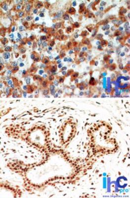
- Immunohistochemistry-Paraffin: Caspase-8 Antibody (90A992) [NB100-56527] - Formalin-fixed, paraffin-embedded human spleen (top) and breast (bottom) stained with Caspase-8 antibody at 4 ug/ml. Localization can be cytoplasmic and nuclear. Cancer/normal adjacent tissue array was used for this test. Staining of formalin-fixed tissues is enhanced by boiling tissue sections in 10 mM sodium citrate buffer, pH 6.0 for 10-20 min followed by cooling at RT for 20 min.
Supportive validation
- Submitted by
- Novus Biologicals (provider)
- Main image
- Experimental details
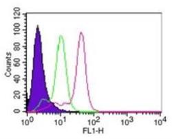
- Flow Cytometry: Caspase-8 Antibody (90A992) [NB100-56527] - Flow cytometric analysis of Caspase-8 in HeLa cells using 0.1 ug of Caspase-8 antibody. Shaded histogram represents cells without antibody; green represents isotype control; red represents Caspase-8 antibody. Goat anti-mouse IgG-FITC secondary antibody was used for this test. IC-Flow (Intracellular Staining Flow Cytometry Kit) was used to fix and prepare the cells for staining.
