Explore
 Validate
 Learn
Learn Western blot
Antibody data
- Antibody Data
- Antigen structure
- References [0]
- Comments [0]
- Validations
- Western blot [2]
- Immunocytochemistry [1]
- Immunoprecipitation [1]
- Immunohistochemistry [2]
- Flow cytometry [1]
Submit
Validation data
Reference
Comment
Report error
- Product number
- MA5-56476 - Provider product page

- Provider
- Invitrogen Antibodies
- Product name
- Caspase 8 Recombinant Rabbit Monoclonal Antibody (PSH04-77)
- Antibody type
- Monoclonal
- Antigen
- Synthetic peptide
- Reactivity
- Human
- Host
- Rabbit
- Isotype
- IgG
- Antibody clone number
- PSH04-77
- Vial size
- 100 μL
- Concentration
- 1 mg/mL
- Storage
- Store at 4°C short term. For long term storage, store at -20°C, avoiding freeze/thaw cycles.
No comments: Submit comment
Supportive validation
- Submitted by
- Invitrogen Antibodies (provider)
- Main image
- Experimental details
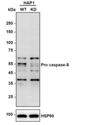
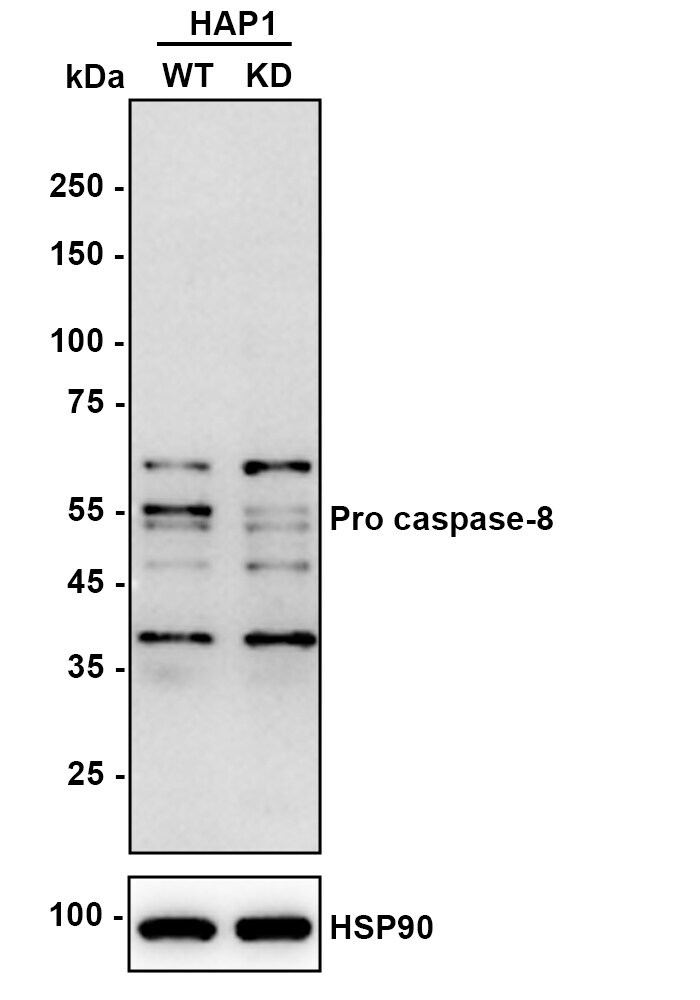
- Western blot analysis of Caspase 8 in Lane 1: HAP1-parental cell lysate, Lane 2: HAP1-Caspase-8 KD cell lysate. (Lysates/Proteins at 10 µg/Lane). Samples were incubated with Caspase 8 Recombinant Monoclonal Antibody (Product # MA5-56476) using a dilution of 1:1,000 Proteins were transferred to a PVDF membrane and blocked with 5% NFDM/TBST (1 hr, room temperature). Followed by using Goat Anti-Rabbit IgG-HRP Secondary Antibody at dilution 1:50,000 (1hr, room temp). Predicted band size: 55 kDa; Observed band size: 54,55 kDa; Exposure time: 180 seconds.
- Submitted by
- Invitrogen Antibodies (provider)
- Main image
- Experimental details
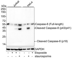
- Western blot analysis of Caspase 8 in Lane 1: Jurkat cell lysate, Lane 2: Jurkat treated with 25μM Etoposide for 5 hours cell lysate, Lane 3: Jurkat treated with 25μM Etoposide for 16 hours cell lysate, Lane 4: Jurkat treated with 1μM Staurosporine for 3 hours cell lysate, Lane 5: HeLa cell lysate, Lane 6: HeLa treated with 1μM Staurosporine for 3 hours cell lysate, Lane 7: HeLa treated with 100μM Etoposide for 4 hours cell lysate. (Lysates/Proteins at 30 µg/Lane). Samples were incubated with Caspase 8 Recombinant Monoclonal Antibody (Product # MA5-56476) using a dilution of 1:2,000 Proteins were transferred to a PVDF membrane and blocked with 5% NFDM/TBST (1 hr, room temperature). Followed by using Goat Anti-Rabbit IgG-HRP Secondary Antibody at dilution 1:50,000 (1hr, room temp). Predicted band size: 55 kDa; Observed band size: 18-55 kDa; Exposure time: 3 minutes.

Supportive validation
- Submitted by
- Invitrogen Antibodies (provider)
- Main image
- Experimental details
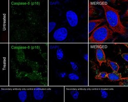
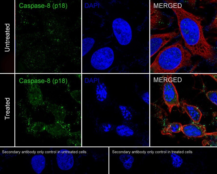
- Immunocytochemistry analysis of Caspase 8 with HeLa cells . Cells were fixed in 4% paraformaldehyde (20 min, room temp), permeabilized with 0.1% Triton X-100 in PBS (5 min, room temp), then blocked with 1% BSA in 10% negative goat serum (1 hr, room temp). Next they were incubated with Caspase 8 Recombinant Monoclonal Antibody (Product # MA5-56476)at a dilution of 1:100 in 1% BSA in PBST overnight at 4 ℃. Followed by Goat Anti-Rabbit IgG H&L at a dilution of 1:1,000. DAPI was used to stain the cell nuclei (blue).
Supportive validation
- Submitted by
- Invitrogen Antibodies (provider)
- Main image
- Experimental details
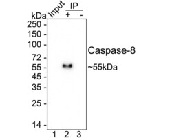
- Caspase 8 was immunoprecipitated from 0.2 mg HeLa treated with 1μM Staurosporine for 3 hours cell lysate with Caspase 8 Recombinant Monoclonal Antibody (Product # MA5-56476) at 2 µg/25 µl agarose. Western blot was performed from the immunoprecipitate using Caspase 8 Recombinant Monoclonal Antibody (Product # MA5-56476) at 1:1,000 dilution. Followed by Anti-Rabbit IgG for IP Nano-secondary antibody at 1:5,000 dilution (1 hr, room temperature). Lane 1: HeLa treated with 1μM Staurosporine for 3 hours cell lysate; Lane 2: (MA5-56476) IP in HeLa treated with 1μM Staurosporine for 3 hours cell lysate; Lane 3: Rabbit IgG in HeLa treated with 1μM Staurosporine for 3 hours cell lysate. Blocking/Dilution buffer: 5% NFDM/TBST. Exposure time: 1 minute 28 seconds.

Supportive validation
- Submitted by
- Invitrogen Antibodies (provider)
- Main image
- Experimental details
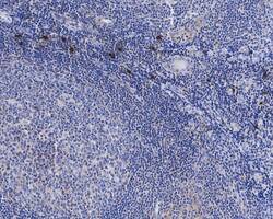
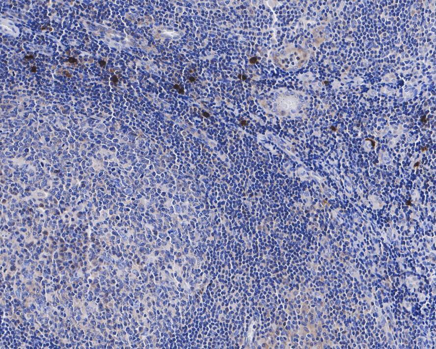
- Immunohistochemistry analysis of Caspase 8 with paraffin-embedded human tonsil tissue. The tissues were blocked in 1% BSA (20 min, room temp), washed with ddH2O and PBS, incubated with Caspase 8 Recombinant Monoclonal Antibody (Product # MA5-56476) at a dilution of 1:1,000 (1 hr, room temp).
- Submitted by
- Invitrogen Antibodies (provider)
- Main image
- Experimental details
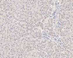
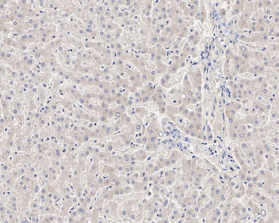
- Immunohistochemistry analysis of Caspase 8 with paraffin-embedded human liver tissue. The tissues were blocked in 1% BSA (20 min, room temp), washed with ddH2O and PBS, incubated with Caspase 8 Recombinant Monoclonal Antibody (Product # MA5-56476) at a dilution of 1:200 (1 hr, room temp).
Supportive validation
- Submitted by
- Invitrogen Antibodies (provider)
- Main image
- Experimental details
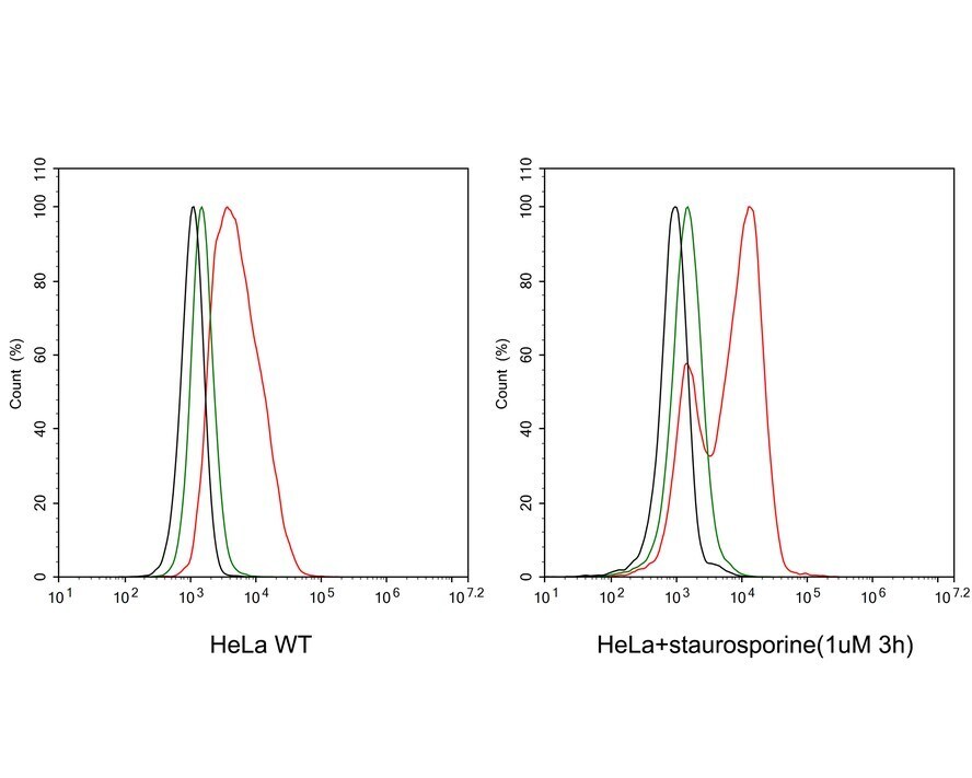
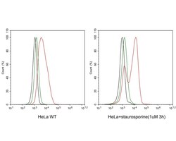
- Flow cytometry analysis of Caspase 8 with HeLa cells. The cells were fixed, permeabilized and incubated with Caspase 8 Recombinant Monoclonal Antibody (Product # MA5-56476) at a dilution of 1μg/mL (red) and compared with Rabbit IgG Isotype Control (green). Followed by iFluor™ 488 conjugate-Goat anti-Rabbit IgG Secondary antibody at 1:1,000 dilution (30 min, 4℃).