 Explore
 Validate
 Learn
Learn Western blot
Antibody data
- Antibody Data
- Antigen structure
- References [0]
- Comments [0]
- Validations
- Western blot [2]
- Immunohistochemistry [2]
- Flow cytometry [1]
Submit
Validation data
Reference
Comment
Report error
- Product number
- SM7015P - Provider product page

- Provider
- Acris Antibodies GmbH
- Proper citation
- Acris Antibodies GmbH Cat#SM7015P, RRID:AB_973848
- Product name
- anti Caspase-8
- Antibody type
- Monoclonal
- Antigen
- Amino acids PVETDSEEQP of Human Caspase-8
- Reactivity
- Human, Simian
- Host
- Mouse
- Isotype
- IgG
- Antibody clone number
- 90A992
- Vial size
- 0.1 mg
- Concentration
- 0.5 mg/ml
No comments: Submit comment
Supportive validation
- Submitted by
- Acris Antibodies GmbH (provider)
- Main image

- Experimental details
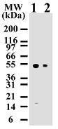
- Western blot analysis for human Caspase-8 using Jurkat lysates with SM7015P at 2 µg/ml (lane 1) and 0.5 µg/ml (lane 2). SM7015P only detects 55 kDa Caspase-8 in Jurkat cells.
- Submitted by
- Acris Antibodies GmbH (provider)
- Main image

- Experimental details
- Western blot analysis of Caspase-8 in Jurkat cells using SM7015P at 1 µg/ml. Cells were treated with 2 µM staurosporine for different time periods. Caspase-8 activation is detected in western blots by the presence of Caspase-8 cleavage fragments. The antibody detected both pro (full-length) and active (cleaved) Caspase-8, depending on the treatment time points. A basal level of endogenously cleaved Caspase-8 can be see in untreated Jurkat cells. HRP conjugated Goat anti-Mouse Ig secondary antibody and PicoTect ECL substrate solution were used for this test.

Supportive validation
- Submitted by
- Acris Antibodies GmbH (provider)
- Main image

- Experimental details
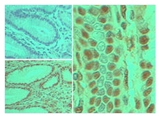
- Immunohistochemical analysis of Caspase-8 in Formalin-Fixed, Paraffin-Embedded Human stomach tumor tissue using an Isotype Control (top left) and SM7015P (bottom left, right) at 5 µg/ml.
- Submitted by
- Acris Antibodies GmbH (provider)
- Main image
- Experimental details
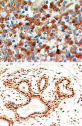
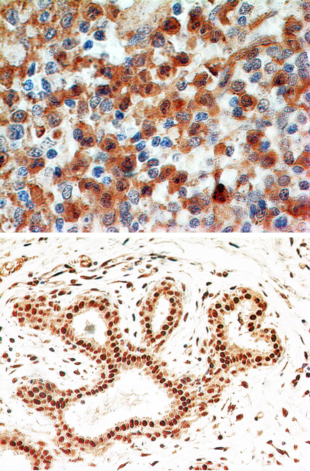
- Formalin-fixed, paraffin-embedded human spleen (top) and breast (bottom) stained with Caspase-8 antibody (Cat.-No.: SM7015P)at 4 µg/ml. Localization can be cytoplasmic and nuclear. Cancer/normal adjacent tissue array was used for this test.Staining of formalin-fixed tissues is enhanced by boiling tissue sections in 10 mM sodium citrate buffer, pH 6.0 for 10-20 min followed by cooling at RT for 20 min.
Supportive validation
- Submitted by
- Acris Antibodies GmbH (provider)
- Main image
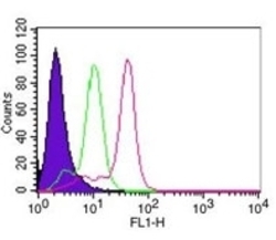
- Experimental details
- Flow Cytometric analysis of Caspase-8 in HeLa cells using 0.1 µg of SM7015P. Shaded histogram represents cells without antibody. Green represents Isotype Control. Red represents anti-Caspase-8 antibody. Goat anti-Mouse IgG-FITC secondary antibody was used for this test. IC-Flow (Intracellular Staining Flow Cytometry Kit) was used to fix and prepare the cells for staining.
