Explore
 Validate
 Learn
Learn Western blot
Antibody data
- Antibody Data
- Antigen structure
- References [2]
- Comments [0]
- Validations
- Western blot [2]
- Immunohistochemistry [1]
- Other assay [1]
Submit
Validation data
Reference
Comment
Report error
- Product number
- 44-1355G - Provider product page

- Provider
- Invitrogen Antibodies
- Product name
- Phospho-Btk (Tyr551) Polyclonal Antibody
- Antibody type
- Polyclonal
- Antigen
- Synthetic peptide
- Reactivity
- Human
- Host
- Rabbit
- Isotype
- IgG
- Vial size
- 100 µL
- Storage
- -20°C
Submitted references Exploration of novel heterofused 1,2,4-triazine derivative in colorectal cancer.
Simultaneous use of erythropoietin and LFM-A13 as a new therapeutic approach for colorectal cancer.
Hermanowicz JM, Szymanowska A, Sieklucka B, Czarnomysy R, Pawlak K, Bielawska A, Bielawski K, Kalafut J, Przybyszewska A, Surazynski A, Rivero-Muller A, Mojzych M, Pawlak D
Journal of enzyme inhibition and medicinal chemistry 2021 Dec;36(1):535-548
Journal of enzyme inhibition and medicinal chemistry 2021 Dec;36(1):535-548
Simultaneous use of erythropoietin and LFM-A13 as a new therapeutic approach for colorectal cancer.
Tankiewicz-Kwedlo A, Hermanowicz JM, Domaniewski T, Pawlak K, Rusak M, Pryczynicz A, Surazynski A, Kaminski T, Kazberuk A, Pawlak D
British journal of pharmacology 2018 Mar;175(5):743-762
British journal of pharmacology 2018 Mar;175(5):743-762
No comments: Submit comment
Supportive validation
- Submitted by
- Invitrogen Antibodies (provider)
- Main image
- Experimental details
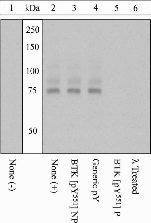
- Western blot analysis of lysates prepared from EB1 cells using rabbit anti-BTK (pY551) antibody (Product # 44-1355G). Cells left untreated (lane 1) or treated with hydrogen peroxide (lanes 2-5) were resolved on a 10 percent polyacrylamide gel and transferred to PVDF. The membrane was treated with either no peptide (lane 2), the non-phosphopeptide corresponding to the phosphopeptide immunogen (lane 3), a generic phosphotyrosine containing peptide (lane 4), or the phosphopeptide immunogen (lane 5). Prior to incubation with the primary antibody, lane 6 was treated with Lambda phosphatase.
- Submitted by
- Invitrogen Antibodies (provider)
- Main image

- Experimental details
- Western blot analysis of BTK (pY551) was performed by loading 20 µg of Daudi (lane1), Daudi treated for 1 hr with 100 uM of Pervanadate (lane2), Jurkat (lane3), Jurkat treated for 1 hr with 100 uM of Pervanadate (lane4), Ramos (lane5) and Ramos treated for 1 hr with 100 uM of Pervanadate (lane6), cell lysate using Novex® NuPAGE® 4-12 % Bis-Tris gel (Product # NP0322BOX), XCell SureLock Electrophoresis System (Product # EI0002), Novex® Sharp Pre-Stained Protein Standard (LC5800), and iBlot® 2 Dry Blotting System (IB21001). Proteins were transferred to a nitrocellulose membrane and blocked with 5 % skim milk at 4°C overnight. BTK (pY551) was detected at ~ 76 kDa using BTK (pY551) Rabbit Polyclonal Antibody (Product # 44-1355G) at 1:1000 dilution in 5 % skim milk for 3 hours at room temperature on a rocking platform. Goat Anti-Rabbit IgG - HRP Secondary Antibody (G21234) at 1:5000 dilution was used and chemiluminescent detection was performed using Pierce™ ECL Western Blotting Substrate (Product # 32106).

Supportive validation
- Submitted by
- Invitrogen Antibodies (provider)
- Main image
- Experimental details
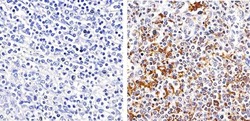
- Immunohistochemistry analysis of Phospho-BTK (pTyr551) showing staining in the cytoplasm of paraffin-embedded human spleen tissue (right) compared to a negative control without primary antibody (left). To expose target proteins, antigen retrieval was performed using 10mM sodium citrate (pH 6.0), microwaved for 8-15 min. Following antigen retrieval, tissues were blocked in 3% H2O2-methanol for 15 min at room temperature, washed with ddH2O and PBS, and then probed with a Phospho-BTK (pTyr551) polyclonal antibody (Product # 44-1355G) diluted in 3% BSA-PBS at a dilution of 1:100 overnight at 4ºC in a humidified chamber. Tissues were washed extensively in PBST and detection was performed using an HRP-conjugated secondary antibody followed by colorimetric detection using a DAB kit. Tissues were counterstained with hematoxylin and dehydrated with ethanol and xylene to prep for mounting.

Supportive validation
- Submitted by
- Invitrogen Antibodies (provider)
- Main image

- Experimental details
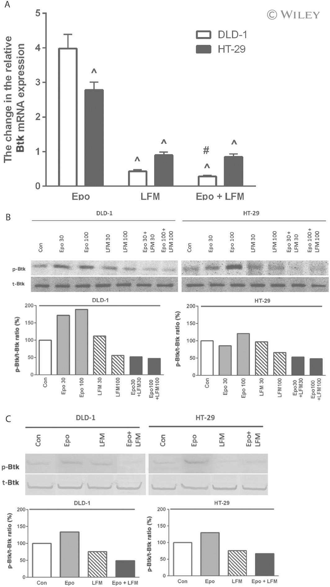
- 2 Btk mRNA levels in DLD-1 and HT-29 cells treated with Epo, LFM-A13 (LFM) and their combination. Results are presented as means +- SD, n = 6. ^ P < 0.05 (vs. Epo), # P < 0.05 (vs. LFM-A13) (A). Phosphorylated Btk (p-Btk) and total Btk (t-Btk) expression as determined by Western blot in DLD-1 and HT-29 cells treated with Epo (30, 100 IU*mL -1 ), LFM-A13 (LFM 30, 100 muM) and their combination for 48 h (Btk) and 5 min (p-Btk). Samples used for electrophoresis consisted of 20 mug of protein from six pooled independent cell extracts ( n = 6). Band staining was quantified by densitometry. Quantitative analysis of the p-Btk/t-Btk ratio, expressed as a percentage of the control level (B). Phosphorylated Btk (p-Btk) and total Btk (t-Btk) expression as determined by Western blot in DLD-1 and HT-29 cells treated with the most effective concentration of Epo (100 IU*mL -1 ), LFM-A13 (LFM 100 muM) and their combination for 48 h (Btk) and 5 min (p-Btk). Samples used for electrophoresis consisted of 20 mug of protein from six pooled independent cell extracts ( n = 6). Band staining was quantified by densitometry. Quantitative analysis of the p-Btk/t-Btk ratio, expressed as a percentage of the control level (C).