 Explore
 Validate
 Learn
Learn Western blot
 Immunocytochemistry
Antibody data
- Antibody Data
- Antigen structure
- References [0]
- Comments [0]
- Validations
- Western blot [1]
- Immunohistochemistry [2]
- Flow cytometry [2]
Submit
Validation data
Reference
Comment
Report error
- Product number
- NBP2-59677 - Provider product page

- Provider
- Novus Biologicals
- Product name
- Armenian Hamster Monoclonal Mre11 Antibody
- Antibody type
- Monoclonal
- Description
- Protein A or G purified.
- Reactivity
- Human, Mouse
- Isotype
- IgG
- Vial size
- 0.1 mg
- Concentration
- 1.0 mg/ml
- Storage
- Store at 4C short term. Aliquot and store at -20C long term. Avoid freeze-thaw cycles.
No comments: Submit comment
Supportive validation
- Submitted by
- Novus Biologicals (provider)
- Main image
- Experimental details
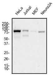
- Western Blot: Mre11 Antibody (15B8.1E7.6) [NBP2-59677] - Total protein from human HeLa, Jurkat, and mouse MEF and Neuro2A cell lines was separated on a 12% gel by SDS-PAGE, transferred to PVDF membrane and blocked in 5% non-fat milk in TBST. The membrane was probed with 1.0 ug/ml anti-Mre11 in block buffer and detected with an anti-Armenian Hamster HRP secondary antibody using chemiluminescence.

Supportive validation
- Submitted by
- Novus Biologicals (provider)
- Main image

- Experimental details
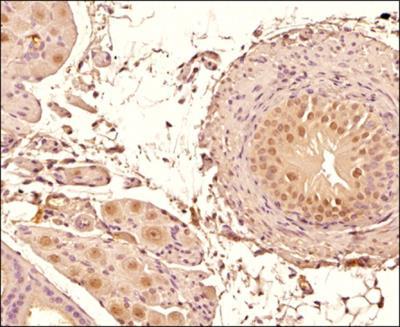
- Immunohistochemistry-Paraffin: Mre11 Antibody (15B8.1E7.6) [NBP2-59677] - IHC analysis of a formalin fixed paraffin embedded (FFPE) tissue section of mouse prostate using 1:500 dilution of Mre11 antibody (clone 15B8.1E7.6). The signal was developed using HRP-DAB indirect detection method and the sections were counterstained using hematoxylin staining. This Mre11 antibody generated a strong nuclear positivity with considerable mild to moderate cytoplasmic staining in the glandular epithelial cells. Select cells depicted MRE11 foci which reflects towards the presence of DNA damage in those cells.
- Submitted by
- Novus Biologicals (provider)
- Main image
- Experimental details
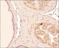
- Immunohistochemistry-Paraffin: Mre11 Antibody (15B8.1E7.6) [NBP2-59677] - IHC analysis of a formalin fixed paraffin embedded (FFPE) tissue section of mouse prostate tissue section using 1:500 dilution of Mre11 antibody (clone 15B8.1E7.6). The signal was developed using HRP-DAB indirect detection method and the sections were counterstained using hematoxylin staining. This Mre11 antibody generated a strong nuclear with mild to moderate cytoplasmic staining in the glandular epithelial cells.

Supportive validation
- Submitted by
- Novus Biologicals (provider)
- Main image
- Experimental details
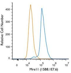
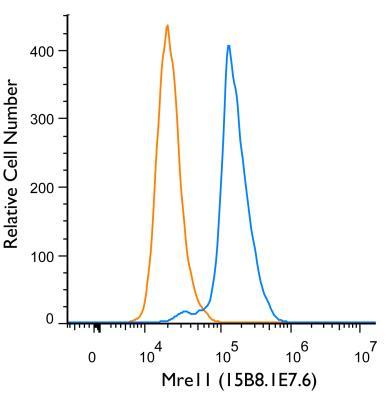
- Flow (Intracellular): Mre11 Antibody (15B8.1E7.6) [NBP2-59677] - An intracellular stain was performed on HeLa Cells with Mre11 (15B8.1E7.6) antibody NBP2-59677 (blue). Unstained cells are shown in orange. Cells were fixed with 4% paraformaldehyde, following fixation, cells were permeabilized with 0.1% saponin. Cells were incubated in an antibody dilution of 1 ug/mL for 30 minutes at room temperature, followed by armenian hamster IgG Alexa Flour 488-conjugated secondary.
- Submitted by
- Novus Biologicals (provider)
- Main image
- Experimental details
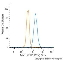
- Flow Cytometry: Mre11 Antibody (15B8.1E7.6) [NBP2-59677] - An intracellular stain was performed on A431 cells with Mre11 [15B8.1E7.6] Antibody NBP2-59677B (blue) and a matched isotype control (orange). Both antibodies were conjugated to Biotin. Cells were fixed with 4% PFA and then permeabilized with 0.1% saponin. Cells were incubated in an antibody dilution of 2.5 ug/mL for 30 minutes at room temperature, followed by Streptavidin - R-Phycoerythrin Protein (2012-1000, Novus Biologicals).
