 Explore
 Validate
 Learn
Learn Western blot
 Immunocytochemistry
Antibody data
- Antibody Data
- Antigen structure
- References [3]
- Comments [0]
- Validations
- Immunocytochemistry [1]
- Immunohistochemistry [26]
- Other assay [1]
Submit
Validation data
Reference
Comment
Report error
- Product number
- PA5-104853 - Provider product page

- Provider
- Invitrogen Antibodies
- Product name
- Phospho-PI3K p85 alpha (Tyr607) Polyclonal Antibody
- Antibody type
- Polyclonal
- Antigen
- Synthetic peptide
- Description
- Antibody detects endogenous levels of PI3K p85 alpha only when phosphorylated at Tyrosine 607.
- Reactivity
- Human, Mouse, Rat, Porcine
- Host
- Rabbit
- Isotype
- IgG
- Vial size
- 100 μL
- Concentration
- 1 mg/mL
- Storage
- -20°C
Submitted references Oxidation of Hemoglobin Drives a Proatherogenic Polarization of Macrophages in Human Atherosclerosis.
Olfactory Bulb Proteomics Reveals Widespread Proteostatic Disturbances in Mixed Dementia and Guides for Potential Serum Biomarkers to Discriminate Alzheimer Disease and Mixed Dementia Phenotypes.
Kaempferol Inhibits Zearalenone-Induced Oxidative Stress and Apoptosis via the PI3K/Akt-Mediated Nrf2 Signaling Pathway: In Vitro and In Vivo Studies.
Potor L, Hendrik Z, Patsalos A, Katona É, Méhes G, Póliska S, Csősz É, Kalló G, Komáromi I, Combi Z, Posta N, Sikura KÉ, Pethő D, Oros M, Vereb G, Tóth C, Gergely P, Nagy L, Balla G, Balla J
Antioxidants & redox signaling 2021 Oct 20;35(12):917-950
Antioxidants & redox signaling 2021 Oct 20;35(12):917-950
Olfactory Bulb Proteomics Reveals Widespread Proteostatic Disturbances in Mixed Dementia and Guides for Potential Serum Biomarkers to Discriminate Alzheimer Disease and Mixed Dementia Phenotypes.
Lachén-Montes M, Íñigo-Marco I, Cartas-Cejudo P, Fernández-Irigoyen J, Santamaría E
Journal of personalized medicine 2021 Jun 3;11(6)
Journal of personalized medicine 2021 Jun 3;11(6)
Kaempferol Inhibits Zearalenone-Induced Oxidative Stress and Apoptosis via the PI3K/Akt-Mediated Nrf2 Signaling Pathway: In Vitro and In Vivo Studies.
Rajendran P, Ammar RB, Al-Saeedi FJ, Mohamed ME, ElNaggar MA, Al-Ramadan SY, Bekhet GM, Soliman AM
International journal of molecular sciences 2020 Dec 28;22(1)
International journal of molecular sciences 2020 Dec 28;22(1)
No comments: Submit comment
Supportive validation
- Submitted by
- Invitrogen Antibodies (provider)
- Main image
- Experimental details
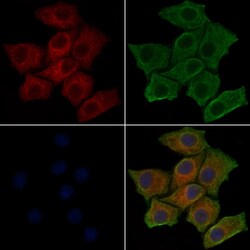
- Immunocytochemistry analysis of Phospho-PI3K p85 alpha (Tyr607) in HepG2 cells (30 min of 4 µM Forskolin treatment). The samples were fixed with PFA and permeabilized in 0.1% Triton X-100, then blocked in 10% serum for 45 minutes at 25°C. Samples were incubated with Phospho-PI3K p85 alpha (Tyr607) polyclonal antibody (Product # PA5-104853) and mouse anti-beta tubulin antibody for 1 hour at 37°C. An AlexaFluor594 conjugated goat anti-rabbit IgG(H+L) (Red) and an AlexaFluor488 conjugated goat anti-mouse IgG(H+L) (Green) were used as the secondary antibodies. The nuclear counter stain is DAPI (blue).
Supportive validation
- Submitted by
- Invitrogen Antibodies (provider)
- Main image
- Experimental details
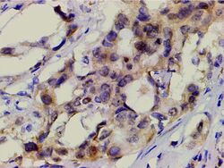
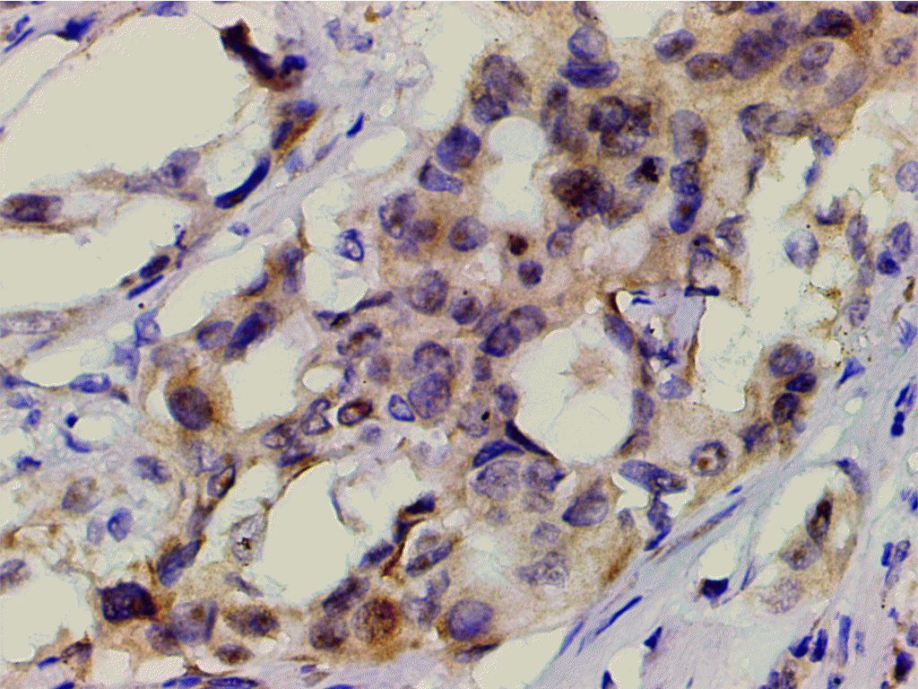
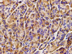
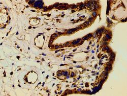
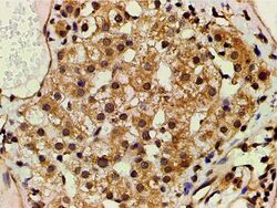
- Immunohistochemistry analysis of paraffin-embedded Phospho-PI3K p85 alpha (Tyr607) in human lung cancer tissue sections. Antigen retrieval was performed using citrate buffer. Samples were blocked with blocking buffer (1.5 hr, 22°C), incubated with Phospho-PI3K p85 alpha (Tyr607) polyclonal antibody (Product # PA5-104853) using a dilution of 1:200 (1.5 hr, 22°C), followed by HRP conjugated goat anti-rabbit.
- Submitted by
- Invitrogen Antibodies (provider)
- Main image

- Experimental details
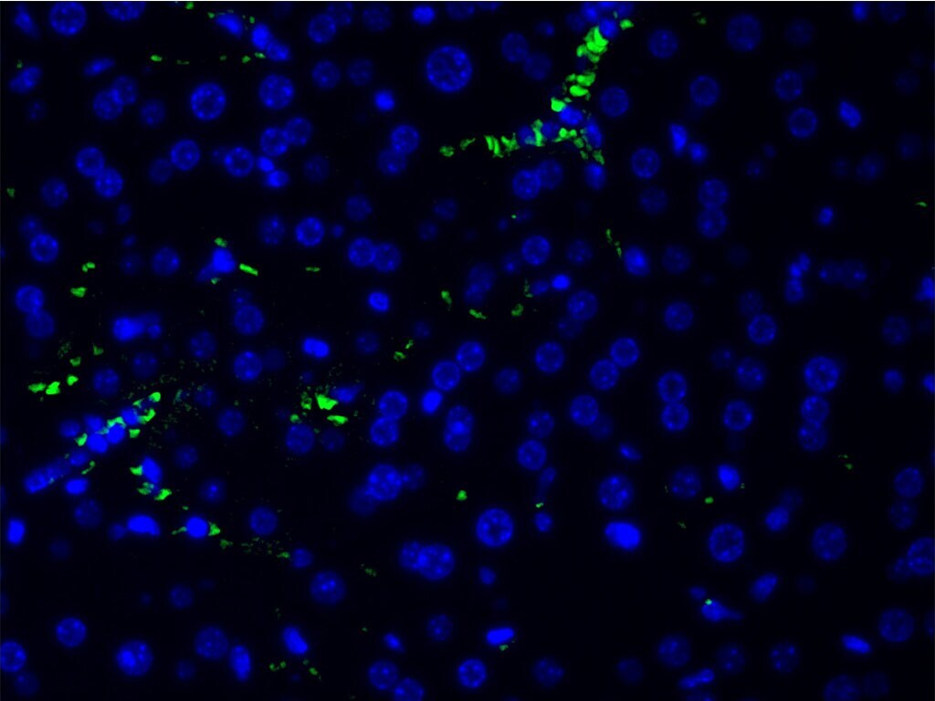
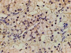
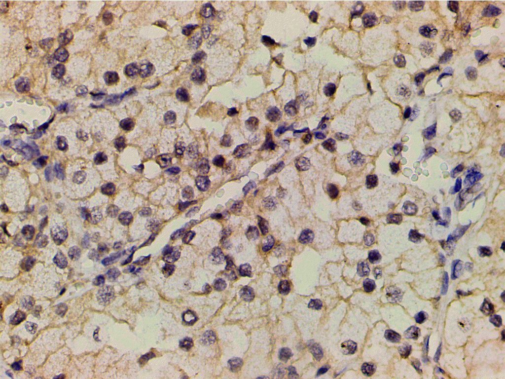
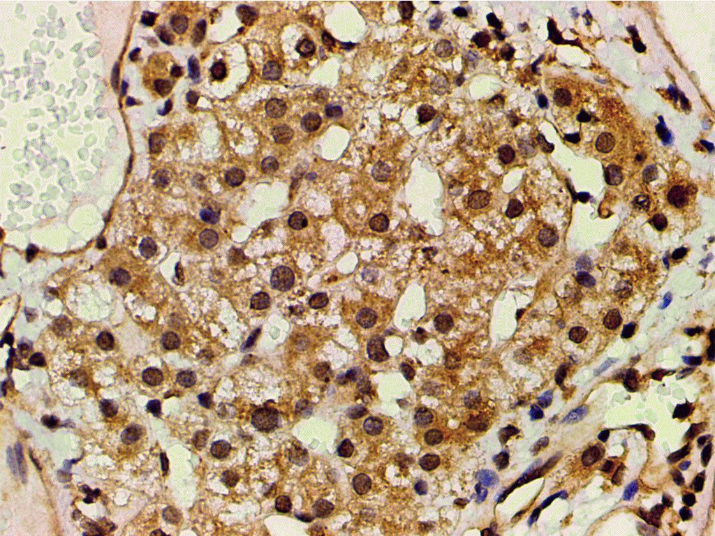
- Immunohistochemistry analysis of Phospho-PI3K p85 alpha (Tyr607) in frozen human liver tissue section. Samples were incubated with Phospho-PI3K p85 alpha (Tyr607) polyclonal antibody (Product # PA5-104853) using a dilution of 1:200 in BSA for 1 hour followed by Alexa Fluor 488®-conjugated Goat anti-rabbit secondary antibody.
- Submitted by
- Invitrogen Antibodies (provider)
- Main image
- Experimental details
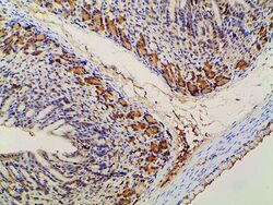
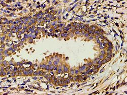
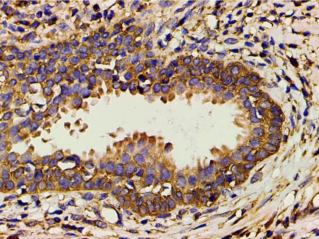
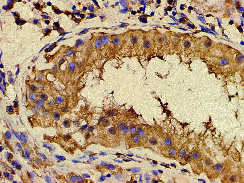
- Immunohistochemistry analysis of paraffin-embedded Phospho-PI3K p85 alpha (Tyr607) in rat uterine tissue sections. Antigen retrieval was performed using citrate buffer. Samples were blocked with blocking buffer (1.5 hr, 22°C), incubated with Phospho-PI3K p85 alpha (Tyr607) polyclonal antibody (Product # PA5-104853) using a dilution of 1:100 (1.5 hr, 22°C), followed by HRP conjugated goat anti-rabbit.
- Submitted by
- Invitrogen Antibodies (provider)
- Main image
- Experimental details
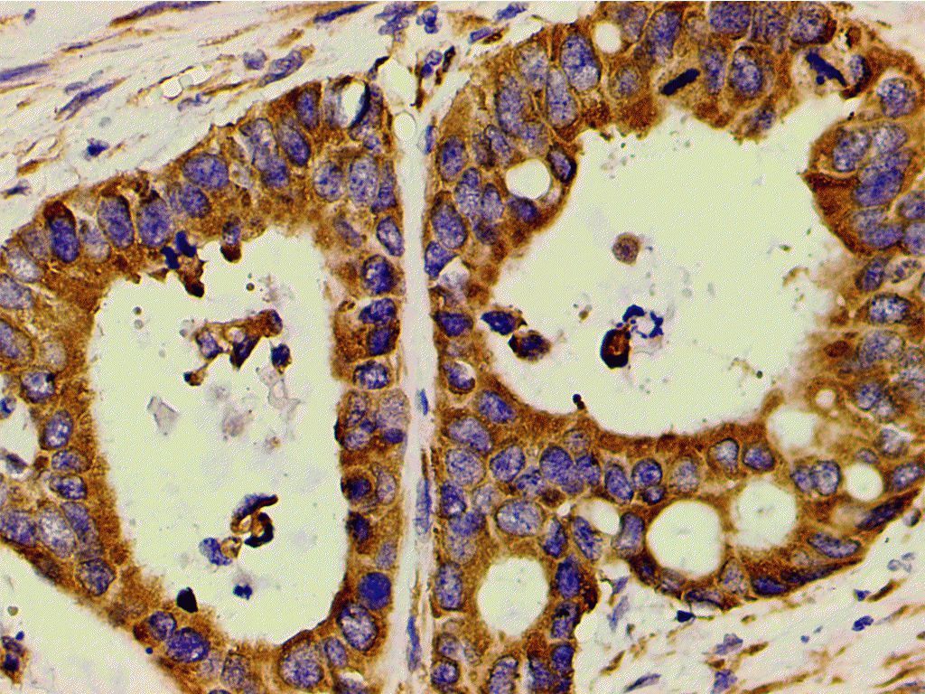
- Immunohistochemistry analysis of paraffin-embedded Phospho-PI3K p85 alpha (Tyr607) in rat Intestinal tissue sections. Antigen retrieval was performed using citrate buffer. Samples were blocked with blocking buffer (1.5 hr, 22°C), incubated with Phospho-PI3K p85 alpha (Tyr607) polyclonal antibody (Product # PA5-104853) using a dilution of 1:100 (1.5 hr, 22°C), followed by HRP conjugated goat anti-rabbit.

- Submitted by
- Invitrogen Antibodies (provider)
- Main image
- Experimental details
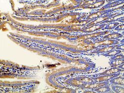
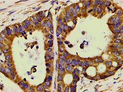
- Immunohistochemistry analysis of paraffin-embedded Phospho-PI3K p85 alpha (Tyr607) in human duodenum tissue sections. Antigen retrieval was performed using citrate buffer. Samples were blocked with blocking buffer (1.5 hr, 22°C), incubated with Phospho-PI3K p85 alpha (Tyr607) polyclonal antibody (Product # PA5-104853) using a dilution of 1:200 (1.5 hr, 22°C), followed by HRP conjugated goat anti-rabbit.

- Submitted by
- Invitrogen Antibodies (provider)
- Main image
- Experimental details
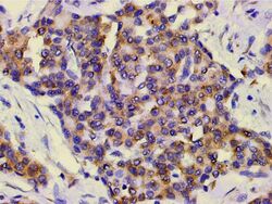
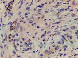
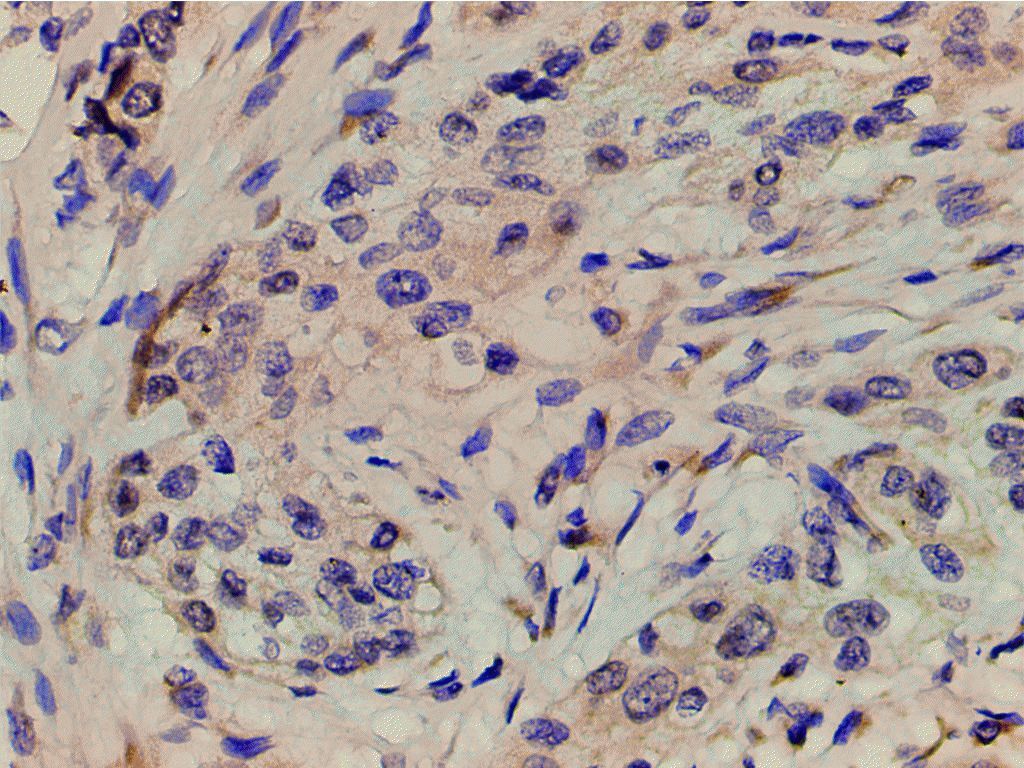
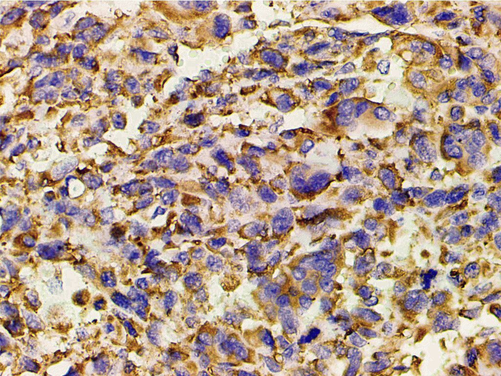
- Immunohistochemistry analysis of paraffin-embedded Phospho-PI3K p85 alpha (Tyr607) in human bladder cancer tissue sections. Antigen retrieval was performed using citrate buffer. Samples were blocked with blocking buffer (1.5 hr, 22°C), incubated with Phospho-PI3K p85 alpha (Tyr607) polyclonal antibody (Product # PA5-104853) using a dilution of 1:200 (1.5 hr, 22°C), followed by HRP conjugated goat anti-rabbit.
- Submitted by
- Invitrogen Antibodies (provider)
- Main image
- Experimental details
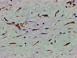
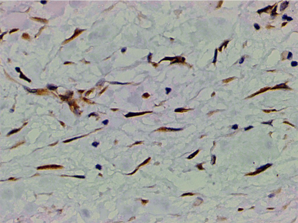
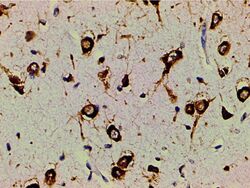
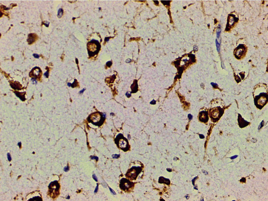
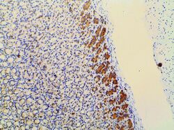
- Immunohistochemistry analysis of paraffin-embedded Phospho-PI3K p85 alpha (Tyr607) in human brain tissue sections. Antigen retrieval was performed using citrate buffer. Samples were blocked with blocking buffer (1.5 hr, 22°C), incubated with Phospho-PI3K p85 alpha (Tyr607) polyclonal antibody (Product # PA5-104853) using a dilution of 1:200 (1.5 hr, 22°C), followed by HRP conjugated goat anti-rabbit.
- Submitted by
- Invitrogen Antibodies (provider)
- Main image
- Experimental details
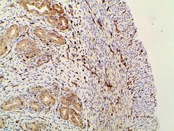
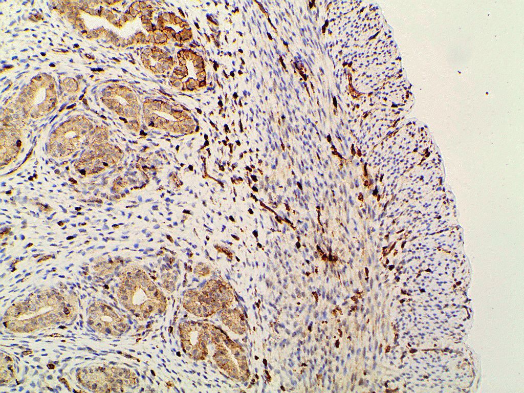
- Immunohistochemistry analysis of paraffin-embedded Phospho-PI3K p85 alpha (Tyr607) in human colon cancer tissue sections. Antigen retrieval was performed using citrate buffer. Samples were blocked with blocking buffer (1.5 hr, 22°C), incubated with Phospho-PI3K p85 alpha (Tyr607) polyclonal antibody (Product # PA5-104853) using a dilution of 1:200 (1.5 hr, 22°C), followed by HRP conjugated goat anti-rabbit.
- Submitted by
- Invitrogen Antibodies (provider)
- Main image
- Experimental details
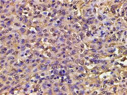
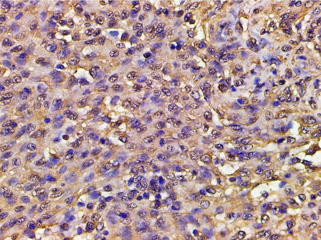
- Immunohistochemistry analysis of paraffin-embedded Phospho-PI3K p85 alpha (Tyr607) in human liver cancer tissue sections. Antigen retrieval was performed using citrate buffer. Samples were blocked with blocking buffer (1.5 hr, 22°C), incubated with Phospho-PI3K p85 alpha (Tyr607) polyclonal antibody (Product # PA5-104853) using a dilution of 1:200 (1.5 hr, 22°C), followed by HRP conjugated goat anti-rabbit.

- Submitted by
- Invitrogen Antibodies (provider)
- Main image
- Experimental details
- Immunohistochemistry analysis of paraffin-embedded Phospho-PI3K p85 alpha (Tyr607) in human esophageal carcinoma tissue sections. Antigen retrieval was performed using citrate buffer. Samples were blocked with blocking buffer (1.5 hr, 22°C), incubated with Phospho-PI3K p85 alpha (Tyr607) polyclonal antibody (Product # PA5-104853) using a dilution of 1:200 (1.5 hr, 22°C), followed by HRP conjugated goat anti-rabbit.
- Submitted by
- Invitrogen Antibodies (provider)
- Main image
- Experimental details
- Immunohistochemistry analysis of paraffin-embedded Phospho-PI3K p85 alpha (Tyr607) in rat ovarian tissue sections. Antigen retrieval was performed using citrate buffer. Samples were blocked with blocking buffer (1.5 hr, 22°C), incubated with Phospho-PI3K p85 alpha (Tyr607) polyclonal antibody (Product # PA5-104853) using a dilution of 1:100 (1.5 hr, 22°C), followed by HRP conjugated goat anti-rabbit.
- Submitted by
- Invitrogen Antibodies (provider)
- Main image
- Experimental details
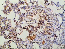
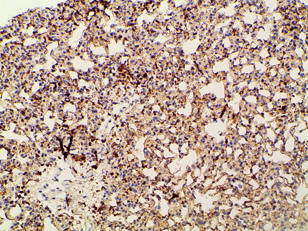
- Immunohistochemistry analysis of paraffin-embedded Phospho-PI3K p85 alpha (Tyr607) in rat lung tissue sections. Antigen retrieval was performed using citrate buffer. Samples were blocked with blocking buffer (1.5 hr, 22°C), incubated with Phospho-PI3K p85 alpha (Tyr607) polyclonal antibody (Product # PA5-104853) using a dilution of 1:100 (1.5 hr, 22°C), followed by HRP conjugated goat anti-rabbit.
- Submitted by
- Invitrogen Antibodies (provider)
- Main image
- Experimental details
- Immunohistochemistry analysis of paraffin-embedded Phospho-PI3K p85 alpha (Tyr607) in rat kidney tissue sections. Antigen retrieval was performed using citrate buffer. Samples were blocked with blocking buffer (1.5 hr, 22°C), incubated with Phospho-PI3K p85 alpha (Tyr607) polyclonal antibody (Product # PA5-104853) using a dilution of 1:100 (1.5 hr, 22°C), followed by HRP conjugated goat anti-rabbit.

- Submitted by
- Invitrogen Antibodies (provider)
- Main image

- Experimental details
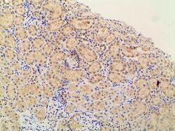
- Immunohistochemistry analysis of paraffin-embedded Phospho-PI3K p85 alpha (Tyr607) in mouse pancreatic tissue sections. Antigen retrieval was performed using citrate buffer. Samples were blocked with blocking buffer (1.5 hr, 22°C), incubated with Phospho-PI3K p85 alpha (Tyr607) polyclonal antibody (Product # PA5-104853) using a dilution of 1:100 (1.5 hr, 22°C), followed by HRP conjugated goat anti-rabbit.
- Submitted by
- Invitrogen Antibodies (provider)
- Main image
- Experimental details
- Immunohistochemistry analysis of paraffin-embedded Phospho-PI3K p85 alpha (Tyr607) in rat gastric tissue sections. Antigen retrieval was performed using citrate buffer. Samples were blocked with blocking buffer (1.5 hr, 22°C), incubated with Phospho-PI3K p85 alpha (Tyr607) polyclonal antibody (Product # PA5-104853) using a dilution of 1:100 (1.5 hr, 22°C), followed by HRP conjugated goat anti-rabbit.

- Submitted by
- Invitrogen Antibodies (provider)
- Main image
- Experimental details
- Immunohistochemistry analysis of paraffin-embedded Phospho-PI3K p85 alpha (Tyr607) in mouse lung tissue sections. Antigen retrieval was performed using citrate buffer. Samples were blocked with blocking buffer (1.5 hr, 22°C), incubated with Phospho-PI3K p85 alpha (Tyr607) polyclonal antibody (Product # PA5-104853) using a dilution of 1:100 (1.5 hr, 22°C), followed by HRP conjugated goat anti-rabbit.
- Submitted by
- Invitrogen Antibodies (provider)
- Main image
- Experimental details
- Immunohistochemistry analysis of paraffin-embedded Phospho-PI3K p85 alpha (Tyr607) in mouse gastric tissue sections. Antigen retrieval was performed using citrate buffer. Samples were blocked with blocking buffer (1.5 hr, 22°C), incubated with Phospho-PI3K p85 alpha (Tyr607) polyclonal antibody (Product # PA5-104853) using a dilution of 1:100 (1.5 hr, 22°C), followed by HRP conjugated goat anti-rabbit.

- Submitted by
- Invitrogen Antibodies (provider)
- Main image
- Experimental details
- Immunohistochemistry analysis of paraffin-embedded Phospho-PI3K p85 alpha (Tyr607) in human renal clear cell carcinoma tissue sections. Antigen retrieval was performed using citrate buffer. Samples were blocked with blocking buffer (1.5 hr, 22°C), incubated with Phospho-PI3K p85 alpha (Tyr607) polyclonal antibody (Product # PA5-104853) using a dilution of 1:200 (1.5 hr, 22°C), followed by HRP conjugated goat anti-rabbit.
- Submitted by
- Invitrogen Antibodies (provider)
- Main image
- Experimental details
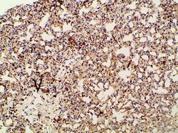
- Immunohistochemistry analysis of paraffin-embedded Phospho-PI3K p85 alpha (Tyr607) in human meningeal carcinomatosis(MC) tissue sections. Antigen retrieval was performed using citrate buffer. Samples were blocked with blocking buffer (1.5 hr, 22°C), incubated with Phospho-PI3K p85 alpha (Tyr607) polyclonal antibody (Product # PA5-104853) using a dilution of 1:200 (1.5 hr, 22°C), followed by HRP conjugated goat anti-rabbit.
- Submitted by
- Invitrogen Antibodies (provider)
- Main image
- Experimental details
- Immunohistochemistry analysis of paraffin-embedded Phospho-PI3K p85 alpha (Tyr607) in human myosarcoma tissue sections. Antigen retrieval was performed using citrate buffer. Samples were blocked with blocking buffer (1.5 hr, 22°C), incubated with Phospho-PI3K p85 alpha (Tyr607) polyclonal antibody (Product # PA5-104853) using a dilution of 1:200 (1.5 hr, 22°C), followed by HRP conjugated goat anti-rabbit.
- Submitted by
- Invitrogen Antibodies (provider)
- Main image

- Experimental details
- Immunohistochemistry analysis of paraffin-embedded Phospho-PI3K p85 alpha (Tyr607) in human osteosarcoma tissue sections. Antigen retrieval was performed using citrate buffer. Samples were blocked with blocking buffer (1.5 hr, 22°C), incubated with Phospho-PI3K p85 alpha (Tyr607) polyclonal antibody (Product # PA5-104853) using a dilution of 1:200 (1.5 hr, 22°C), followed by HRP conjugated goat anti-rabbit.
- Submitted by
- Invitrogen Antibodies (provider)
- Main image
- Experimental details
- Immunohistochemistry analysis of paraffin-embedded Phospho-PI3K p85 alpha (Tyr607) in human placenta tissue sections. Antigen retrieval was performed using citrate buffer. Samples were blocked with blocking buffer (1.5 hr, 22°C), incubated with Phospho-PI3K p85 alpha (Tyr607) polyclonal antibody (Product # PA5-104853) using a dilution of 1:200 (1.5 hr, 22°C), followed by HRP conjugated goat anti-rabbit.
- Submitted by
- Invitrogen Antibodies (provider)
- Main image
- Experimental details
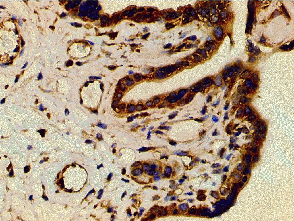
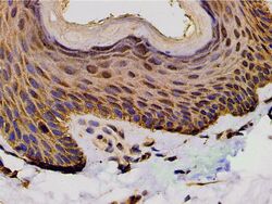
- Immunohistochemistry analysis of paraffin-embedded Phospho-PI3K p85 alpha (Tyr607) in human skin tissue sections. Antigen retrieval was performed using citrate buffer. Samples were blocked with blocking buffer (1.5 hr, 22°C), incubated with Phospho-PI3K p85 alpha (Tyr607) polyclonal antibody (Product # PA5-104853) using a dilution of 1:200 (1.5 hr, 22°C), followed by HRP conjugated goat anti-rabbit.

- Submitted by
- Invitrogen Antibodies (provider)
- Main image
- Experimental details
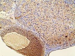
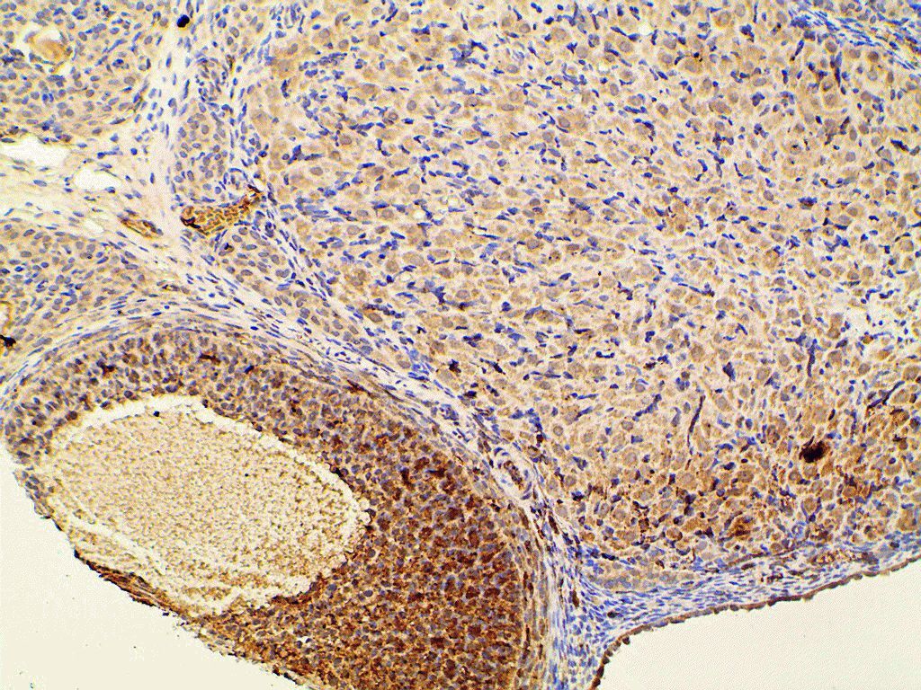
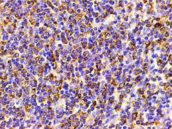
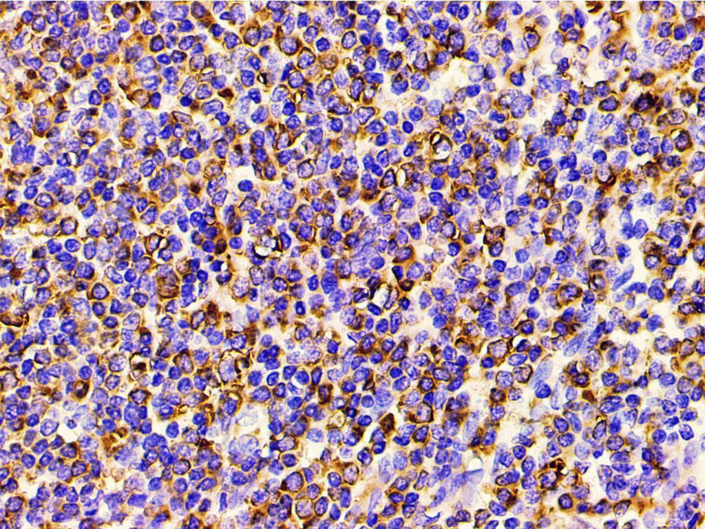
- Immunohistochemistry analysis of paraffin-embedded Phospho-PI3K p85 alpha (Tyr607) in human spleen tissue sections. Antigen retrieval was performed using citrate buffer. Samples were blocked with blocking buffer (1.5 hr, 22°C), incubated with Phospho-PI3K p85 alpha (Tyr607) polyclonal antibody (Product # PA5-104853) using a dilution of 1:200 (1.5 hr, 22°C), followed by HRP conjugated goat anti-rabbit.
- Submitted by
- Invitrogen Antibodies (provider)
- Main image
- Experimental details
- Immunohistochemistry analysis of paraffin-embedded Phospho-PI3K p85 alpha (Tyr607) in human vascular cancer tissue sections. Antigen retrieval was performed using citrate buffer. Samples were blocked with blocking buffer (1.5 hr, 22°C), incubated with Phospho-PI3K p85 alpha (Tyr607) polyclonal antibody (Product # PA5-104853) using a dilution of 1:200 (1.5 hr, 22°C), followed by HRP conjugated goat anti-rabbit.
- Submitted by
- Invitrogen Antibodies (provider)
- Main image

- Experimental details
- Immunohistochemistry analysis of paraffin-embedded Phospho-PI3K p85 alpha (Tyr607) in human seminoma tissue sections. Antigen retrieval was performed using citrate buffer. Samples were blocked with blocking buffer (1.5 hr, 22°C), incubated with Phospho-PI3K p85 alpha (Tyr607) polyclonal antibody (Product # PA5-104853) using a dilution of 1:200 (1.5 hr, 22°C), followed by HRP conjugated goat anti-rabbit.
Supportive validation
- Submitted by
- Invitrogen Antibodies (provider)
- Main image
- Experimental details
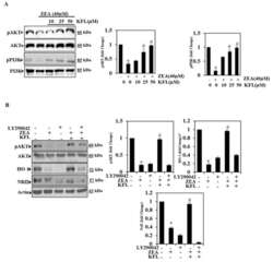
- Figure 4 KFL triggers the PI3K/Akt pathway in ZEA-induced HepG2 cells. ( A ) Cells were treated with KFL (10, 25 and 50 uM) followed by ZEA (40 uM) for 24 h. After the treatment, whole-cell lysates were exposed to Western blotting with anti-pPI3K and anti-pAkt antibodies. Total PI3K and Akt levels were measured as loading controls. ( B ) Cells were pre-treated with a PI3K/Akt inhibitor (LY294002, 30 muM) for 2 h, followed by KFL (50 muM) and/or ZEA (40 muM) for 24 h. Western blot was performed to detect the pAkt, HO-1 and Nrf2 levels by anti-pAkt, anti-HO-1 and anti-Nrf2 abs. Data are represented as the mean +- SD of triplicate values ( n = 3), and * p < 0.05 represents noteworthy discrepancies compared with the control. # p < 0.05 represents significant variations compared with the ZEA alone and KFL with ZEA treatment groups.
