 Explore
 Validate
 Learn
Learn Western blot
 Immunocytochemistry
Antibody data
- Antibody Data
- Antigen structure
- References [5]
- Comments [0]
- Validations
- Immunocytochemistry [2]
- Immunohistochemistry [1]
- Flow cytometry [1]
- Other assay [3]
Submit
Validation data
Reference
Comment
Report error
- Product number
- AHO1272 - Provider product page

- Provider
- Invitrogen Antibodies
- Product name
- FAK Monoclonal Antibody (34Q36)
- Antibody type
- Monoclonal
- Antigen
- Recombinant protein fragment
- Reactivity
- Human, Mouse, Rat
- Host
- Mouse
- Isotype
- IgG
- Antibody clone number
- 34Q36
- Vial size
- 100 μg
- Concentration
- 0.5 mg/mL
- Storage
- -20°C
Submitted references Peruvoside is a novel Src inhibitor that suppresses NSCLC cell growth and motility by downregulating multiple Src-EGFR-related pathways.
Identification of TENM4 as a Novel Cancer Stem Cell-Associated Molecule and Potential Target in Triple Negative Breast Cancer.
Nup93 regulates breast tumor growth by modulating cell proliferation and actin cytoskeleton remodeling.
Human brain microvascular endothelial cell pairs model tissue-level blood-brain barrier function.
Tumor endothelial cells promote metastasis and cancer stem cell-like phenotype through elevated Epiregulin in esophageal cancer.
Lai Y, Chang H, Chen H, Chang G, Chen JJ
American journal of cancer research 2022;12(6):2576-2593
American journal of cancer research 2022;12(6):2576-2593
Identification of TENM4 as a Novel Cancer Stem Cell-Associated Molecule and Potential Target in Triple Negative Breast Cancer.
Ruiu R, Barutello G, Arigoni M, Riccardo F, Conti L, Peppino G, Annaratone L, Marchiò C, Mengozzi G, Calogero RA, Cavallo F, Quaglino E
Cancers 2021 Feb 20;13(4)
Cancers 2021 Feb 20;13(4)
Nup93 regulates breast tumor growth by modulating cell proliferation and actin cytoskeleton remodeling.
Bersini S, Lytle NK, Schulte R, Huang L, Wahl GM, Hetzer MW
Life science alliance 2020 Jan;3(1)
Life science alliance 2020 Jan;3(1)
Human brain microvascular endothelial cell pairs model tissue-level blood-brain barrier function.
O'Connor BB, Grevesse T, Zimmerman JF, Ardoña HAM, Jimenez JA, Bitounis D, Demokritou P, Parker KK
Integrative biology : quantitative biosciences from nano to macro 2020 Apr 14;12(3):64-79
Integrative biology : quantitative biosciences from nano to macro 2020 Apr 14;12(3):64-79
Tumor endothelial cells promote metastasis and cancer stem cell-like phenotype through elevated Epiregulin in esophageal cancer.
Sun L, Pan J, Yu L, Liu H, Shu X, Sun L, Lou J, Yang Z, Ran Y
American journal of cancer research 2016;6(10):2277-2288
American journal of cancer research 2016;6(10):2277-2288
No comments: Submit comment
Supportive validation
- Submitted by
- Invitrogen Antibodies (provider)
- Main image

- Experimental details
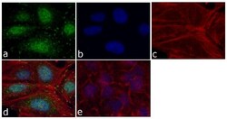
- Immunofluorescent analysis of FAK was done on 70% confluent log phase HeLa cells. The cells were fixed with 4% paraformaldehyde for 15 minutes; permeabilized with 0.25% Triton™ X-100 for 10 minutes followed by blocking with 5% BSA for 1 hour at room temperature. The cells were incubated with FAK Mouse Monoclonal Antibody (Product # AHO1272) at 1:250 dilution in 1% BSA and incubated for 3 hours at room temperature and then labeled with Alexa Fluor 488 Rabbit Anti-Mouse IgG Secondary Antibody (Product # A-11059) at a dilution of 1:400 for 30 minutes at room temperature (Panel a: green). Nuclei (Panel b: blue) were stained with SlowFade® Gold Antifade Mountant with DAPI (Product # S36938). F-actin (Panel c: red) was stained with Alexa Fluor 594 Phalloidin (Product # A12381). Panel d is a merged image showing membrane and cytoplasmic FAK localization. Panel e shows no primary antibody control. The images were captured at 20X magnification.

- Submitted by
- Invitrogen Antibodies (provider)
- Main image
- Experimental details
- Immunofluorescent analysis of FAK was done on 70% confluent log phase HeLa cells. The cells were fixed with 4% paraformaldehyde for 15 minutes; permeabilized with 0.25% Triton™ X-100 for 10 minutes followed by blocking with 5% BSA for 1 hour at room temperature. The cells were incubated with FAK Mouse Monoclonal Antibody (Product # AHO1272) at 1:250 dilution in 1% BSA and incubated for 3 hours at room temperature and then labeled with Alexa Fluor 488 Rabbit Anti-Mouse IgG Secondary Antibody (Product # A-11059) at a dilution of 1:400 for 30 minutes at room temperature (Panel a: green). Nuclei (Panel b: blue) were stained with SlowFade® Gold Antifade Mountant with DAPI (Product # S36938). F-actin (Panel c: red) was stained with Alexa Fluor 594 Phalloidin (Product # A12381). Panel d is a merged image showing membrane and cytoplasmic FAK localization. Panel e shows no primary antibody control. The images were captured at 20X magnification.

Supportive validation
- Submitted by
- Invitrogen Antibodies (provider)
- Main image
- Experimental details
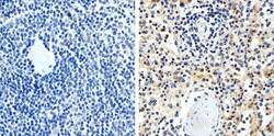
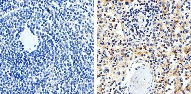
- Immunohistochemistry analysis of FAK (34Q36) showing staining in the cytoplasm and membrane of paraffin-embedded human spleen tissue (right) compared to a negative control without primary antibody (left). To expose target proteins, antigen retrieval was performed using 10 mM sodium citrate (pH 6.0), microwaved for 8-15 min. Following antigen retrieval, tissues were blocked in 3% H2O2-methanol for 15 min at room temperature, washed with ddH2O and PBS, and then probed with FAK (34Q36) monoclonal antibody (Product # AHO1272) diluted in 3% BSA-PBS at a dilution of 1:20 overnight at 4°C in a humidified chamber. Tissues were washed extensively in PBST and detection was performed using a HRP-conjugated secondary antibody followed by colorimetric detection using a DAB kit. Tissues were counterstained with hematoxylin and dehydrated with ethanol and xylene to prep for mounting.
Supportive validation
- Submitted by
- Invitrogen Antibodies (provider)
- Main image
- Experimental details
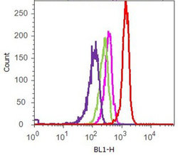
- Flow cytometry analysis of FAK [34Q36] was done on A549 cells. Cells were fixed with 70% ethanol for 10 minutes, permeabilized with 0.25% Tritonª X-100 for 20 minutes, and blocked with 5% BSA for 1 hour at room temperature. Cells were labeled with FAK [34Q36] Mouse Monoclonal Antibody (AHO1272, red histogram) or with mouse isotype control (pink histogram) at 1-3 µg/million cells in 2.5% BSA. After incubation at room temperature for 2-3 hours, the cells were labeled with Alexa Fluor¨ 488 Rabbit Anti-Mouse Secondary Antibody (A11059) at a dilution of 1:400 for 30 minutes at room temperature. The representative 10,000 cells were acquired and analyzed for each sample using an Attune¨ Acoustic Focusing Cytometer. The purple histogram represents unstained control cells and the green histogram represents no-primary-antibody control.

Supportive validation
- Submitted by
- Invitrogen Antibodies (provider)
- Main image

- Experimental details
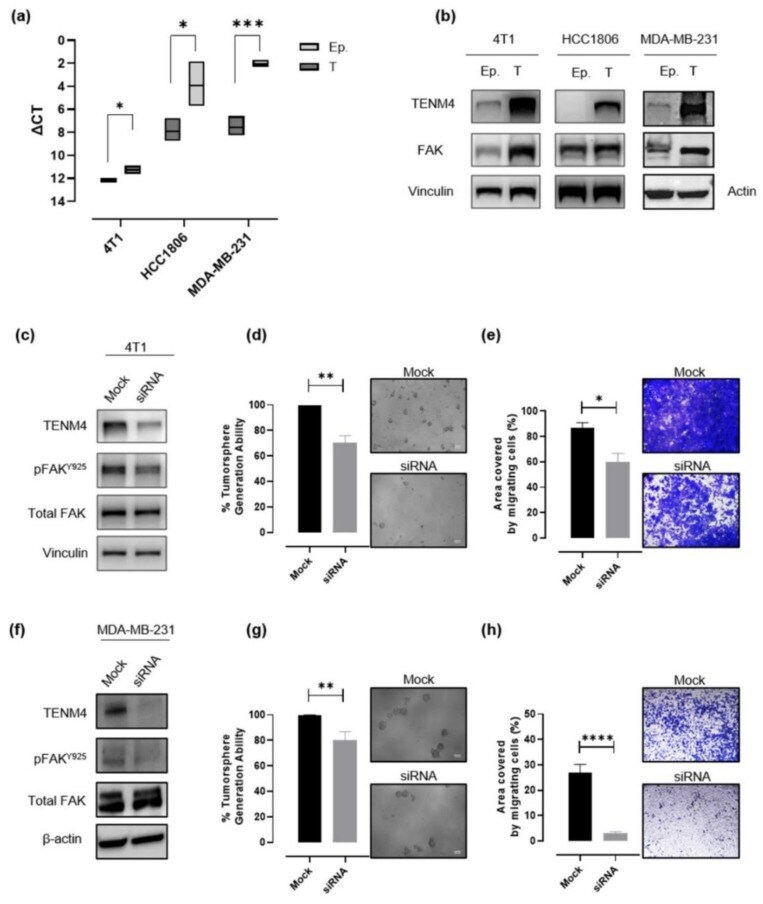
- Figure 4 Evaluation of the role of TENM4 in tumorspheres. ( a ) Semi-quantitative RT-PCR. Boxes represent the DeltaCt (i.e., the difference between the Ct of TENM4 and the Ct of the internal control gene GADPH), comparing epithelial (Ep.) and tumorsphere-derived (T) cells. The inner line of the box corresponds to the mean value, upper and lower edges correspond to higher and lower value, respectively. ( b ) Immunoblot of TENM4, total FAK and loading control protein, vinculin or beta-actin, comparing 4T1, HCC1806 and MDA-MB-231 epithelial (Ep.) and tumorsphere-derived (T) cells. ( c , f ) Immunoblot of TENM4, total FAK, FAK phosphorylated at Y925 and loading control protein, vinculin or beta-actin, comparing 4T1 or MDA-MB-231 epithelial cells, respectively, treated with a pool of TENM4-specific siRNA (siRNA) or a pool of non-targeting siRNA (mock). ( d , g ) Histograms showing the sphere-generation ability and representative pictures of the formed tumorspheres after TENM4 silencing in 4T1 and MDA-MB-231 cells, respectively. The mean values in the mock siRNA-treated condition were used as reference and considered to be 100%. Values of TENM4 silenced cells represent the % of the tumorsphere forming ability respect the mean value of the mock siRNA-treated cells. White scale bar: 100 um. ( e , h ) Effect of TENM4 silencing on 4T1 and MDA-MB-231 cell migratory ability, assessed by using the Transwell migration assay. Histograms showing the mean +- SEM of the percentage of the area
- Submitted by
- Invitrogen Antibodies (provider)
- Main image

- Experimental details
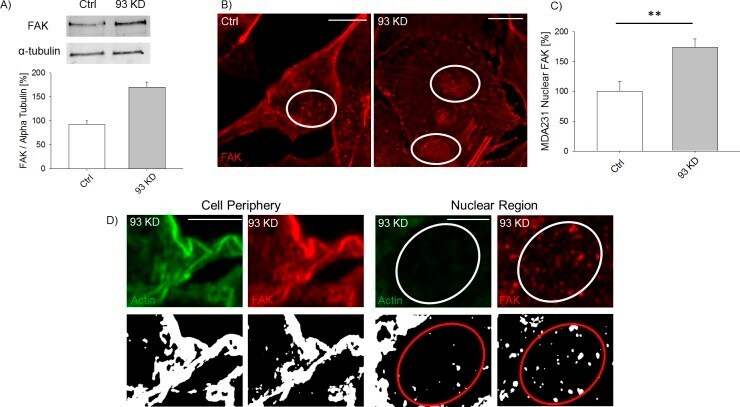
- Figure S3. NUP93 KD increases the overall level of FAK and its nuclear localization. (A) Representative Western blot showing the level of FAK in ctrl and NUP93 KD cells. (B, C) Representative images of FAK localization after NUP93 KD and (C) quantification of its nuclear localization. NUP93 KD FAK staining is from the same image reported in Fig 1A for AC, whereas control FAK staining is from the same image reported in Fig 1E for actin profile plotting. t test with P < 0.05, N >= 6 independent regions, data normalized to ctrl cells. (D) Representative confocal and binary images showing that the actin/FAK co-localization at the cell periphery is lost inside the nucleus of NUP93 KD cells. (B, D) Scale bars: 10 mum (B) and 5 mum (D).
- Submitted by
- Invitrogen Antibodies (provider)
- Main image
- Experimental details
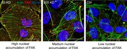
- Figure S6. Altering the Nup93 sub-complex (which includes Nup205) does not affect nucleocytoplasmic transport. Immunofluorescence of FAK in NUP93 KD, NUP205 KD, and ctrl MDA-MB-231 BCCs. Nuclear accumulation of FAK is mainly observed in NUP93 KD cells suggesting that altering the Nup93-Nup205 sub-complex does not significantly affect nucleocytoplasmic transport. The image used for NUP93 KD is also used in Fig 1A . Scale bars: 10 mum.
