Explore
 Validate
 Learn
Learn Western blot
Antibody data
- Antibody Data
- Antigen structure
- References [2]
- Comments [0]
- Validations
- Western blot [2]
- Immunohistochemistry [1]
Submit
Validation data
Reference
Comment
Report error
- Product number
- AF4467 - Provider product page

- Provider
- R&D Systems
- Product name
- Human/Mouse/Rat FAK Antibody
- Antibody type
- Polyclonal
- Description
- Antigen Affinity-purified. Detects human, mouse and rat FAK.
- Reactivity
- Human, Mouse, Rat
- Host
- Sheep
- Conjugate
- Unconjugated
- Antigen sequence
Q05397- Isotype
- IgG
- Vial size
- 100 ug
- Concentration
- LYOPH
- Storage
- Use a manual defrost freezer and avoid repeated freeze-thaw cycles. 12 months from date of receipt, -20 to -70 °C as supplied. 1 month, 2 to 8 °C under sterile conditions after reconstitution. 6 months, -20 to -70 °C under sterile conditions after reconstitution.
Submitted references Focal adhesion kinase coordinates costamere-related JNK signaling with muscle fiber transformation after Achilles tenotomy and tendon reconstruction.
Knee Extensors Muscle Plasticity Over a 5-Years Rehabilitation Process After Open Knee Surgery.
Ferrié C, Kasper S, Wanivenhaus F, Flück M
Experimental and molecular pathology 2019 Jun;108:42-56
Experimental and molecular pathology 2019 Jun;108:42-56
Knee Extensors Muscle Plasticity Over a 5-Years Rehabilitation Process After Open Knee Surgery.
Flück M, Viecelli C, Bapst AM, Kasper S, Valdivieso P, Franchi MV, Ruoss S, Lüthi JM, Bühler M, Claassen H, Hoppeler H, Gerber C
Frontiers in physiology 2018;9:1343
Frontiers in physiology 2018;9:1343
No comments: Submit comment
Supportive validation
- Submitted by
- R&D Systems (provider)
- Main image
- Experimental details
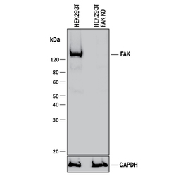
- Western Blot Shows Human FAK Specificity by Using Knockout Cell Line. Western blot shows lysates of HEK293T human embryonic kidney parental cell line and FAK knockout HEK293T cell line (KO). PVDF membrane was probed with 1 µg/mL of Sheep Anti-Human/Mouse/Rat FAK Antigen Affinity-purified Polyclonal Antibody (Catalog # AF4467) followed by HRP-conjugated Anti-Sheep IgG Secondary Antibody (Catalog # HAF016). A specific band was detected for FAK at approximately 135 kDa (as indicated) in the parental HEK293T cell line, but is not detectable in knockout HEK293T cell line. GAPDH (Catalog # AF5718) is shown as a loading control. This experiment was conducted under reducing conditions and using Immunoblot Buffer Group 1.
- Submitted by
- R&D Systems (provider)
- Main image
- Experimental details
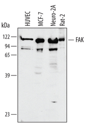
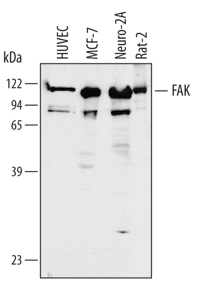
- Detection of Human/Mouse/Rat FAK by Western Blot. Western blot shows lysates of HUVEC human umbilical vein endothelial cells, MCF-7 human breast cancer cell line, Neuro-2A mouse neuroblastoma cell line, and Rat-2 rat embryonic fibroblast cell line. PVDF membrane was probed with 1 µg/mL of Human/Mouse/Rat FAK Antigen Affinity-purified Polyclonal Antibody (Catalog # AF4467) followed by HRP-conjugated Anti-Sheep IgG Secondary Antibody (Catalog # HAF016). A specific band was detected for FAK at approximately 122 kDa (as indicated). This experiment was conducted under reducing conditions and using Immunoblot Buffer Group 1.
Supportive validation
- Submitted by
- R&D Systems (provider)
- Main image
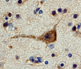
- Experimental details
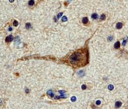
- FAK in Human Brain. FAK was detected in immersion fixed paraffin-embedded sections of human brain (hippocampus) using 3 µg/mL Human/Mouse/Rat FAK Antigen Affinity-purified Polyclonal Antibody (Catalog # AF4467) overnight at 4 °C. Tissue was stained with the Anti-Sheep HRP-DAB Cell & Tissue Staining Kit (brown; Catalog # CTS019) and counterstained with hematoxylin (blue). View our protocol for Chromogenic IHC Staining of Paraffin-embedded Tissue Sections.