Explore
 Validate
 Learn
Learn Western blot
Antibody data
- Antibody Data
- Antigen structure
- References [2]
- Comments [0]
- Validations
- Western blot [1]
- Immunocytochemistry [2]
- Immunoprecipitation [1]
- Immunohistochemistry [1]
- Chromatin Immunoprecipitation [1]
Submit
Validation data
Reference
Comment
Report error
- Product number
- GTX113963 - Provider product page

- Provider
- GeneTex
- Proper citation
- GeneTex Cat#GTX113963, RRID:AB_11168950
- Product name
- UHRF1 antibody
- Antibody type
- Polyclonal
- Reactivity
- Human
- Host
- Rabbit
Submitted references H3K9 methyltransferase G9a negatively regulates UHRF1 transcription during leukemia cell differentiation.
A proteomic characterization of factors enriched at nascent DNA molecules.
Kim KB, Son HJ, Choi S, Hahm JY, Jung H, Baek HJ, Kook H, Hahn Y, Kook H, Seo SB
Nucleic acids research 2015 Apr 20;43(7):3509-23
Nucleic acids research 2015 Apr 20;43(7):3509-23
A proteomic characterization of factors enriched at nascent DNA molecules.
Lopez-Contreras AJ, Ruppen I, Nieto-Soler M, Murga M, Rodriguez-Acebes S, Remeseiro S, Rodrigo-Perez S, Rojas AM, Mendez J, Muñoz J, Fernandez-Capetillo O
Cell reports 2013 Apr 25;3(4):1105-16
Cell reports 2013 Apr 25;3(4):1105-16
No comments: Submit comment
Supportive validation
- Submitted by
- GeneTex (provider)
- Main image

- Experimental details
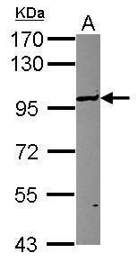
- Sample (30 ?g of whole cell lysate) A: HCT116 7.5% SDS PAGE GTX113963 diluted at 1:1000 The HRP-conjugated anti-rabbit IgG antibody (GTX213110-01) was used to detect the primary antibody.
Supportive validation
- Submitted by
- GeneTex (provider)
- Main image
- Experimental details
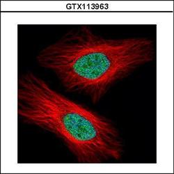
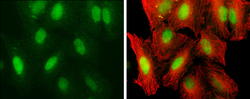
- Confocal immunofluorescence analysis (Olympus FV10i) of paraformaldehyde-fixed HeLa, using UHRF1(GTX113963) antibody (Green) at 1:500 dilution. Alpha-tubulin filaments were labeled with GTX11304 (Red) at 1:2000.
- Submitted by
- GeneTex (provider)
- Main image
- Experimental details
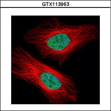
- UHRF1 antibody detects UHRF1 protein at nucleus by immunofluorescent analysis.Sample: A549 cells were fixed in 4% paraformaldehyde at RT for 15 min.Green: UHRF1 protein stained by UHRF1 antibody (GTX113963) diluted at 1:250.Red: alpha Tubulin, a cytoskeleton marker, stained by alpha Tubulin antibody [GT114] (GTX628802) diluted at 1:1000.

Supportive validation
- Submitted by
- GeneTex (provider)
- Main image
- Experimental details
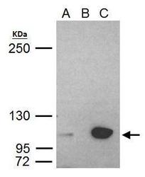
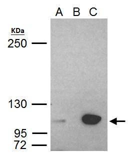
- UHRF1 antibody immunoprecipitates UHRF1 protein in IP experiments. IP Sample: HeLa whole cell lysate/extract A. 40 £gg HeLa whole cell lysate/extract B. Control with 2 £gg of preimmune rabbit IgG C. Immunoprecipitation of UHRF1 protein by 2 £gg of UHRF1 antibody (GTX113963) 5% SDS-PAGE The immunoprecipitated UHRF1 protein was detected by UHRF1 antibody (GTX113963) diluted at 1:1000. EasyBlot anti-rabbit IgG (GTX221666-01) was used as a secondary reagent.
Supportive validation
- Submitted by
- GeneTex (provider)
- Main image

- Experimental details
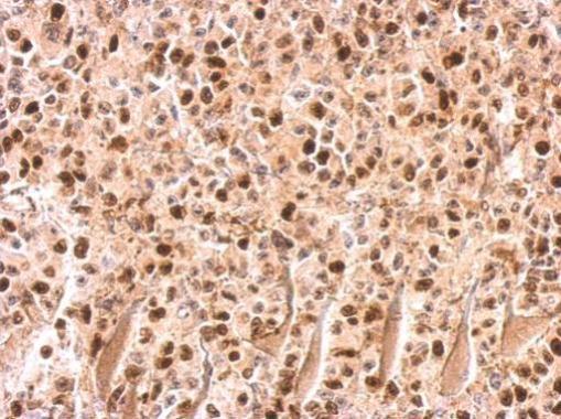
- UHRF1 antibody detects UHRF1 protein at on HBL435 xenograft by immunohistochemical analysis. Sample: Paraffin-embedded HBL435 xenograft. UHRF1 antibody (GTX113963) dilution: 1:500.
Supportive validation
- Submitted by
- GeneTex (provider)
- Main image
- Experimental details
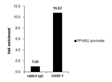
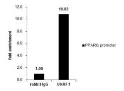
- Cross-linked ChIP was performed with HCT116 chromatin extract and 5 £gg of either control rabbit IgG or anti-UHRF1 antibody. The precipitated DNA was detected by PCR with primer set targeting to PPARG promoter.