Explore
 Validate
 Learn
LearnPA5-27969
antibody from Invitrogen Antibodies
Targeting: UHRF1
FLJ21925, ICBP90, Np95, RNF106, TDRD22
 Western blot
 Immunocytochemistry
Antibody data
- Antibody Data
- Antigen structure
- References [2]
- Comments [0]
- Validations
- Immunocytochemistry [1]
- Immunohistochemistry [1]
- Other assay [2]
Submit
Validation data
Reference
Comment
Report error
- Product number
- PA5-27969 - Provider product page

- Provider
- Invitrogen Antibodies
- Product name
- UHRF1 Polyclonal Antibody
- Antibody type
- Polyclonal
- Antigen
- Recombinant full-length protein
- Description
- Recommended positive controls: HeLa. Predicted reactivity: Bovine (84%). Store product as a concentrated solution. Centrifuge briefly prior to opening the vial.
- Reactivity
- Human, Mouse, Rat
- Host
- Rabbit
- Isotype
- IgG
- Vial size
- 100 μL
- Concentration
- 1 mg/mL
- Storage
- Store at 4°C short term. For long term storage, store at -20°C, avoiding freeze/thaw cycles.
Submitted references MiR-9-1 Suppresses Cell Proliferation and Promotes Apoptosis by Targeting UHRF1 in Lung Cancer.
LASP-1: a nuclear hub for the UHRF1-DNMT1-G9a-Snail1 complex.
Jia CY, Xiang W, Liu JB, Jiang GX, Sun F, Wu JJ, Yang XL, Xin R, Shi Y, Zhang DD, Li W, Zuberi Z, Zhang J, Lu GX, Wang HM, Wang PY, Yu F, Lv ZW, Ma YS, Fu D
Technology in cancer research & treatment 2021 Jan-Dec;20:15330338211041191
Technology in cancer research & treatment 2021 Jan-Dec;20:15330338211041191
LASP-1: a nuclear hub for the UHRF1-DNMT1-G9a-Snail1 complex.
Duvall-Noelle N, Karwandyar A, Richmond A, Raman D
Oncogene 2016 Mar 3;35(9):1122-33
Oncogene 2016 Mar 3;35(9):1122-33
No comments: Submit comment
Supportive validation
- Submitted by
- Invitrogen Antibodies (provider)
- Main image
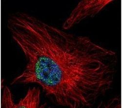
- Experimental details
- Immunofluorescent analysis of UHRF1 in paraformaldehyde-fixed HeLa cells using a UHRF1 polyclonal antibody (Product # PA5-27969) (Green) at a 1:500 dilution. Alpha-tubulin filaments were labeled with Product # PA5-29281 (Red) at a 1:2000.
Supportive validation
- Submitted by
- Invitrogen Antibodies (provider)
- Main image
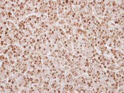
- Experimental details
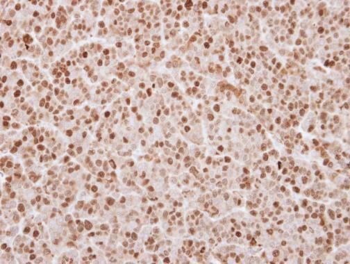
- Immunohistochemical analysis of paraffin-embedded BT483 xenograft, using UHRF1 (Product # PA5-27969) antibody at 1:250 dilution. Antigen Retrieval: Citrate buffer, pH 6.0, 15 min.
Supportive validation
- Submitted by
- Invitrogen Antibodies (provider)
- Main image
- Experimental details
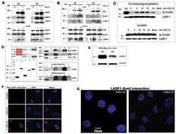
- Fig. 6 LASP-1 associates with epigenetic proteins UHRF1, DNMT1, G9a and Snail1 A) LASP-1 co-immunoprecipitates with UHRF1 and DNMT1. 100 mug of the nuclear extracts from MDA-Bone-Un cells stimulated with vehicle (0 min) or 500 ng /ml of CXCL12 for 15 min were incubated with control mouse monoclonal IgG (Mock) or with mouse anti-LASP-1 antibody. LASP1-bound proteins were eluted and analyzed for endogenous LASP-1 and associated proteins UHRF1 and DNMT1. Bottom panel: 15 mug of total cell lysates from MDA-Bone-Un cells that were stimulated with vehicle (0 min) or CXCL12 for 15 min. LASP-1 lysate blot was obtained after stripping and re-probing for LASP-1 after the detection of Snail1 first. The experiment was repeated twice and the representative blot was quantified and shown. NS - Non-silenced control; KD - LASP-1 knock down. The band intensities of proteins were quantified by using Image J analysis and normalized to their respective non-stimulated control (bands in -CXCL12 lane). The fold change was given above the bands to track the level of the immunoprecipitated proteins with respect to their protein levels in the lysates. B) LASP-1 co-immunoprecipitates with G9a and Snail1. 100 mug of the nuclear extracts from MDA-Bone-Un cells stimulated with vehicle (0 min and Mock) or 500 ng /ml of CXCL12 for 15 min were incubated with control mouse monoclonal IgG (Mock) or with mouse anti-LASP-1 antibody. Bound proteins were eluted and analyzed for endogenous LASP-1 and associated G9a
- Submitted by
- Invitrogen Antibodies (provider)
- Main image
- Experimental details
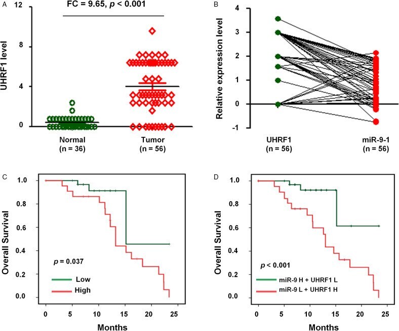
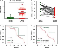
- Figure 6. The expression and clinical significance of miR-9 to 1 and UHRF1 in NSCLC. (A) IHC assay to measure UHRF1 level in 56 NSCLC tissues and 36 adjacently normal lung tissues. (B) The correlation between UHRF1 and miR-9 to 1 level in 56 NSCLC tissues. (C) Kaplan-Meier survival analysis was used to evaluate the prognostic value of UHRF1 expression for OS of 56 NSCLC tissues. (D) Kaplan-Meier survival analysis to evaluate the prognostic value of UHRF1 together with miR-9 to 1 expression in NSCLC for OS. Abbreviations: NSCLC, nonsmall cell lung cancer; miR-9 to 1, microRNA-9 to 1; OS, overall survival; UHRF1, ubiquitin-like containing PHD and RING finger domains 1.