 Explore
 Validate
 Learn
LearnPA5-29884
antibody from Invitrogen Antibodies
Targeting: UHRF1
FLJ21925, ICBP90, Np95, RNF106, TDRD22
 Western blot
 Immunocytochemistry
Antibody data
- Antibody Data
- Antigen structure
- References [2]
- Comments [0]
- Validations
- Immunocytochemistry [5]
- Immunoprecipitation [3]
- Immunohistochemistry [1]
- Chromatin Immunoprecipitation [4]
- Other assay [7]
Submit
Validation data
Reference
Comment
Report error
- Product number
- PA5-29884 - Provider product page

- Provider
- Invitrogen Antibodies
- Product name
- UHRF1 Polyclonal Antibody
- Antibody type
- Polyclonal
- Antigen
- Recombinant full-length protein
- Description
- Recommended positive controls: HCT116, K562. Store product as a concentrated solution. Centrifuge briefly prior to opening the vial.
- Reactivity
- Human, Mouse, Rat
- Host
- Rabbit
- Isotype
- IgG
- Vial size
- 100 μL
- Concentration
- 1.4 mg/mL
- Storage
- Store at 4°C short term. For long term storage, store at -20°C, avoiding freeze/thaw cycles.
Submitted references The microRNA signature of patients with sunitinib failure: regulation of UHRF1 pathways by microRNA-101 in renal cell carcinoma.
Regulation of UHRF1 by dual-strand tumor-suppressor microRNA-145 (miR-145-5p and miR-145-3p): Inhibition of bladder cancer cell aggressiveness.
Goto Y, Kurozumi A, Nohata N, Kojima S, Matsushita R, Yoshino H, Yamazaki K, Ishida Y, Ichikawa T, Naya Y, Seki N
Oncotarget 2016 Sep 13;7(37):59070-59086
Oncotarget 2016 Sep 13;7(37):59070-59086
Regulation of UHRF1 by dual-strand tumor-suppressor microRNA-145 (miR-145-5p and miR-145-3p): Inhibition of bladder cancer cell aggressiveness.
Matsushita R, Yoshino H, Enokida H, Goto Y, Miyamoto K, Yonemori M, Inoguchi S, Nakagawa M, Seki N
Oncotarget 2016 May 10;7(19):28460-87
Oncotarget 2016 May 10;7(19):28460-87
No comments: Submit comment
Supportive validation
- Submitted by
- Invitrogen Antibodies (provider)
- Main image
- Experimental details
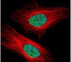
- Immunofluorescent analysis of UHRF1 in paraformaldehyde-fixed HeLa cells using a UHRF1 polyclonal antibody (Product # PA5-29884) (Green) at a 1:500 dilution. Alpha-tubulin filaments were labeled with Product # PA5-29281 (Red) at a 1:2000.
- Submitted by
- Invitrogen Antibodies (provider)
- Main image

- Experimental details
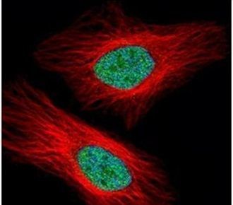
- Immunocytochemistry-Immunofluorescence analysis of UHRF1 was performed in A549 cells fixed in 4% paraformaldehyde at RT for 15 min. Green: UHRF1 Polyclonal Antibody (Product # PA5-29884) diluted at 1:250. Red: alpha Tubulin, a cytoskeleton marker.

- Submitted by
- Invitrogen Antibodies (provider)
- Main image

- Experimental details
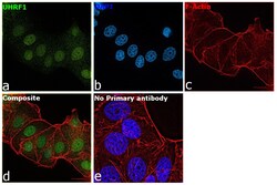
- Immunofluorescence analysis of UHRF1 was performed using 70 confluent log phase MCF7 cells. The cells were fixed with 4% paraformaldehyde for 10 minutes, permeabilized with 0.1% Triton™ X-100 for 15 minutes, and blocked with 2% BSA for 45 minutes at room temperature. The cells were labeled with UHRF1 Polyclonal Antibody (Product # PA5-29884) at 1:100 dilution in 0.1% BSA, incubated at 4 degree celsius overnight and then labeled with Donkey anti-Rabbit IgG (H+L) Highly Cross-Adsorbed Secondary Antibody, Alexa Fluor Plus 488 (Product # A32790), (1:2000 dilution), for 45 minutes at room temperature (Panel a: Green). Nuclei (Panel b:Blue) were stained with ProLong™ Diamond Antifade Mountant with DAPI (Product # P36962). F-actin (Panel c: Red) was stained with Rhodamine Phalloidin (Product # R415, 1:300 dilution). Panel d represents the merged image showing Predominant Nuclear along with faint cytoplasmic localization. Panel e represents control cells with no primary antibody to assess background. The images were captured at 60X magnification.

- Submitted by
- Invitrogen Antibodies (provider)
- Main image
- Experimental details
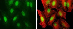
- Immunocytochemistry-Immunofluorescence analysis of UHRF1 was performed in A549 cells fixed in 4% paraformaldehyde at RT for 15 min. Green: UHRF1 Polyclonal Antibody (Product # PA5-29884) diluted at 1:250. Red: alpha Tubulin, a cytoskeleton marker.

- Submitted by
- Invitrogen Antibodies (provider)
- Main image
- Experimental details
- Immunofluorescence analysis of UHRF1 was performed using 70 confluent log phase MCF7 cells. The cells were fixed with 4% paraformaldehyde for 10 minutes, permeabilized with 0.1% Triton™ X-100 for 15 minutes, and blocked with 2% BSA for 45 minutes at room temperature. The cells were labeled with UHRF1 Polyclonal Antibody (Product # PA5-29884) at 1:100 dilution in 0.1% BSA, incubated at 4 degree celsius overnight and then labeled with Donkey anti-Rabbit IgG (H+L) Highly Cross-Adsorbed Secondary Antibody, Alexa Fluor Plus 488 (Product # A32790), (1:2000 dilution), for 45 minutes at room temperature (Panel a: Green). Nuclei (Panel b:Blue) were stained with ProLong™ Diamond Antifade Mountant with DAPI (Product # P36962). F-actin (Panel c: Red) was stained with Rhodamine Phalloidin (Product # R415, 1:300 dilution). Panel d represents the merged image showing Predominant Nuclear along with faint cytoplasmic localization. Panel e represents control cells with no primary antibody to assess background. The images were captured at 60X magnification.

Supportive validation
- Submitted by
- Invitrogen Antibodies (provider)
- Main image
- Experimental details
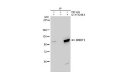
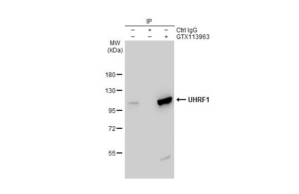
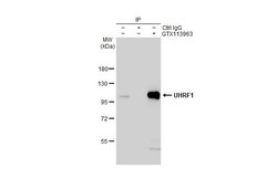
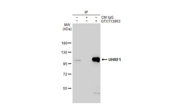
- Immunoprecipitation of UHRF1 protein from HeLa whole cell extract using 5 µg of UHRF1 Polyclonal Antibody (Product # PA5-29884). Western blot analysis was performed using UHRF1 Polyclonal Antibody (Product # PA5-29884).
- Submitted by
- Invitrogen Antibodies (provider)
- Main image

- Experimental details
- Immunoprecipitation of UHRF1 protein from HeLa whole cell extract using 5 µg of UHRF1 Polyclonal Antibody (Product # PA5-29884). Western blot analysis was performed using UHRF1 Polyclonal Antibody (Product # PA5-29884).

- Submitted by
- Invitrogen Antibodies (provider)
- Main image
- Experimental details
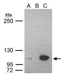
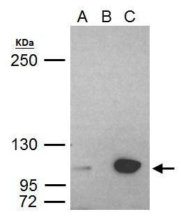
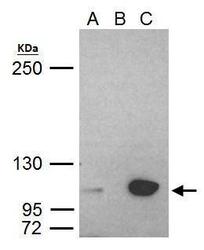
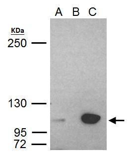
- UHRF1 Polyclonal Antibody immunoprecipitates UHRF1 protein in IP experiments. IP Sample: HeLa whole cell lysate/extract A. 40 µg HeLa whole cell lysate/extract B. Control with 2 µg of preimmune rabbit IgG C. Immunoprecipitation of UHRF1 protein by 2 µg of UHRF1 Polyclonal Antibody (Product # PA5-29884) 5% SDS-PAGE The immunoprecipitated UHRF1 protein was detected by UHRF1 Polyclonal Antibody (Product # PA5-29884) diluted at 1:1,000.
Supportive validation
- Submitted by
- Invitrogen Antibodies (provider)
- Main image
- Experimental details
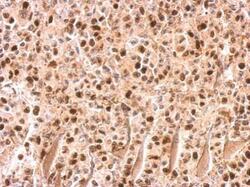
- UHRF1 Polyclonal Antibody detects UHRF1 protein at on HBL435 xenograft by immunohistochemical analysis. Sample: Paraffin-embedded HBL435 xenograft. UHRF1 Polyclonal Antibody (Product # PA5-29884) dilution: 1:500. Antigen Retrieval: EDTA based buffer, pH 8.0, 15 min.
Supportive validation
- Submitted by
- Invitrogen Antibodies (provider)
- Main image

- Experimental details
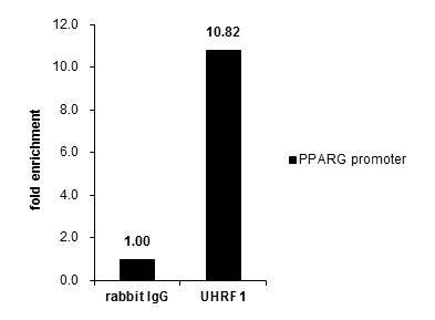
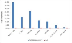
- Cross-linked ChIP was performed with HCT116 chromatin extract and 5 µg of either control rabbit IgG or UHRF1 Polyclonal Antibody (Product # PA5-29884). The precipitated DNA was detected by PCR with primer set targeting to PPARG promoter.
- Submitted by
- Invitrogen Antibodies (provider)
- Main image

- Experimental details
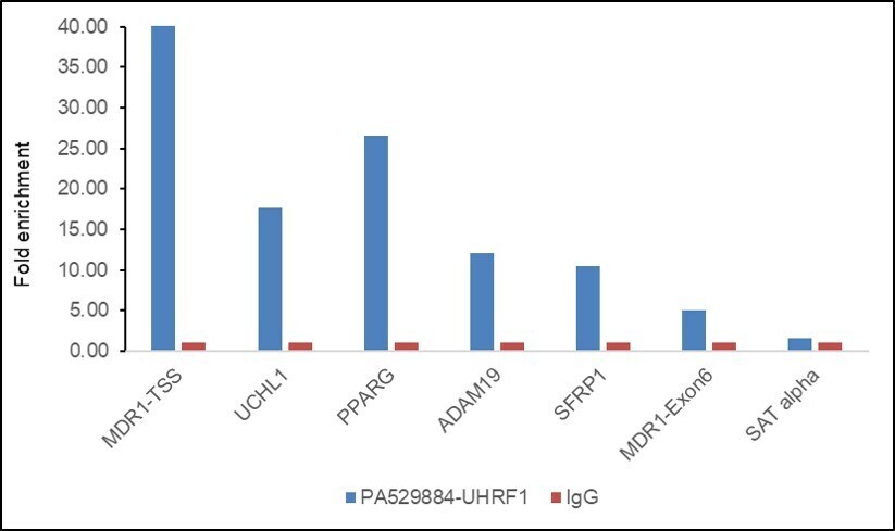
- Chromatin Immunoprecipitation (ChIP) assay of endogenous UHRF1 protein using UHRF1 Antibody: ChIP was performed using UHRF1 Polyclonal Antibody (Product # PA5-29884, 5 µg) on sheared chromatin from HCT 116 cells using the MAGnify ChIP System kit (Product # 49-2024). Normal Rabbit IgG was used as a negative IP control. The purified DNA was analyzed by qPCR using primers binding to MDR1-TSS, UCHL1, PPARG, ADAM19, SFRP1 (active) and MDR1-Exon6 and SAT alpha satellite repeats (Inactive). Data is presented as fold enrichment of the antibody signal versus the negative control IgG using the comparative CT method.

- Submitted by
- Invitrogen Antibodies (provider)
- Main image
- Experimental details
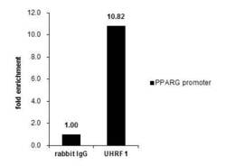
- Cross-linked ChIP was performed with HCT116 chromatin extract and 5 µg of either control rabbit IgG or UHRF1 Polyclonal Antibody (Product # PA5-29884). The precipitated DNA was detected by PCR with primer set targeting to PPARG promoter.

- Submitted by
- Invitrogen Antibodies (provider)
- Main image
- Experimental details
- Chromatin Immunoprecipitation (ChIP) assay of endogenous UHRF1 protein using UHRF1 Antibody: ChIP was performed using UHRF1 Polyclonal Antibody (Product # PA5-29884, 5 µg) on sheared chromatin from HCT 116 cells using the MAGnify ChIP System kit (Product # 49-2024). Normal Rabbit IgG was used as a negative IP control. The purified DNA was analyzed by qPCR using primers binding to MDR1-TSS, UCHL1, PPARG, ADAM19, SFRP1 (active) and MDR1-Exon6 and SAT alpha satellite repeats (Inactive). Data is presented as fold enrichment of the antibody signal versus the negative control IgG using the comparative CT method.
Supportive validation
- Submitted by
- Invitrogen Antibodies (provider)
- Main image
- Experimental details
- Immunoprecipitation of UHRF1 protein from HeLa whole cell extract using 5 µg of UHRF1 Polyclonal Antibody (Product # PA5-29884). Western blot analysis was performed using UHRF1 Polyclonal Antibody (Product # PA5-29884).
- Submitted by
- Invitrogen Antibodies (provider)
- Main image
- Experimental details
- UHRF1 Polyclonal Antibody immunoprecipitates UHRF1 protein in IP experiments. IP Sample: HeLa whole cell lysate/extract A. 40 µg HeLa whole cell lysate/extract B. Control with 2 µg of preimmune rabbit IgG C. Immunoprecipitation of UHRF1 protein by 2 µg of UHRF1 Polyclonal Antibody (Product # PA5-29884) 5% SDS-PAGE The immunoprecipitated UHRF1 protein was detected by UHRF1 Polyclonal Antibody (Product # PA5-29884) diluted at 1:1,000.
- Submitted by
- Invitrogen Antibodies (provider)
- Main image
- Experimental details
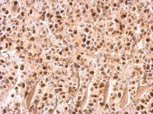
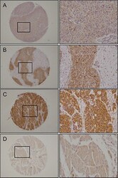
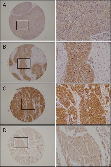
- Figure 9 Immunohistochemical staining of UHRF1 in BC clinical specimens UHRF1 was expressed more strongly in several cancer lesions than in noncancerous tissues. Left panel, original magnification x40; Right panel, original magnification x200. ( A ) Positively stained tumor lesion (High grade, T2bN0M0), ( B ) Positively stained tumor lesion (High grade, T1N0M0), ( C ) Positively stained tumor lesion (Low grade, T3N0M0), ( D ) Negative staining in normal bladder tissue.
- Submitted by
- Invitrogen Antibodies (provider)
- Main image

- Experimental details
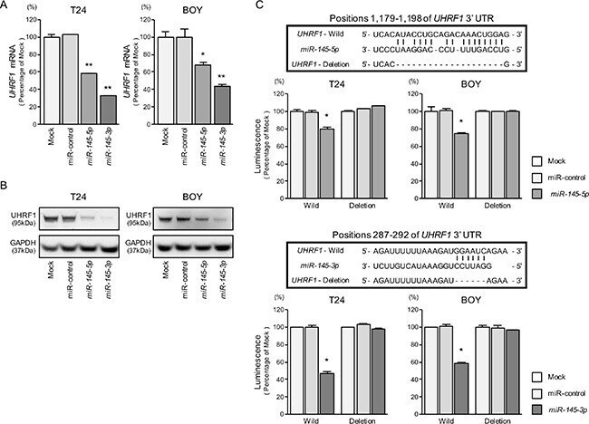
- Figure 4 Direct regulation of UHRF1 by miR-145-5p and miR-145-3p ( A ) UHRF1 mRNA expression was evaluated by qRT-PCR in T24 and BOY 72 hours after transfection with miR-145-5p and miR-145-3p . GUSB was used as an internal control. * P = 0.0036 and ** P < 0.0001. ( B ) UHRF1 protein expression was evaluated by Western blot analyses in T24 and BOY 72-96 hours after transfection with miR-145-5p or miR-145-3p . GAPDH was used as a loading control. ( C ) miR-145-5p and miR-145-3p binding sites in the 3' UTR of UHRF1 mRNA. Dual Luciferase reporter assays using vectors encoding putative miR-145-5p and miR-145-3p target sites of the UHRF 3' UTR (positions 1,179-1,198 and 287-292, respectively) for both wild-type and deleted regions. Normalized data were calculated as ratios of Renilla /firefly luciferase activities. * P < 0.0001.
- Submitted by
- Invitrogen Antibodies (provider)
- Main image

- Experimental details
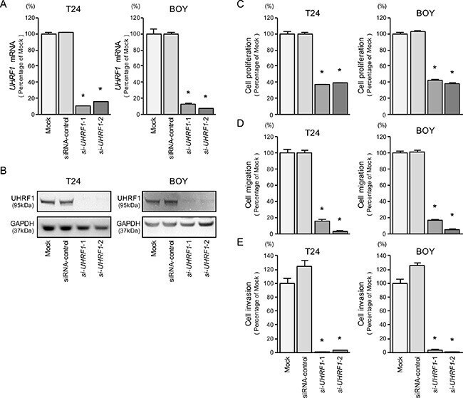
- Figure 5 UHRF1 mRNA and protein expression after si-UHRF1 transfection and effects of UHRF1 silencing in BC cell lines ( A ) UHRF1 mRNA expression was evaluated by qRT-PCR in T24 and BOY 72 hours after transfection with si-UHRF1 -1 and si-UHRF1 -2. GUSB was used as an internal control. ( B ) UHRF1 protein expression was evaluated by Western blot analysis in T24 and BOY 72 - 96 hours after transfection with miR-145-5p or miR-145-3p . GAPDH was used as a loading control. ( C ) Cell proliferation was determined with the XTT assays 72 hours after transfection with 10 nM si-UHRF1 -1 or si-UHRF1 -2. * P < 0.0001. ( D ) Cell migration activity was determined by wound-healing assays. * P < 0.0001. ( E ) Cell invasion activity was determined using Matrigel invasion assays. * P < 0.0001.
- Submitted by
- Invitrogen Antibodies (provider)
- Main image
- Experimental details
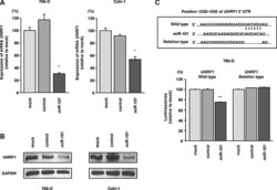
- Figure 3 miR-101 directly downregulated UHRF1 expression in RCC cells ( A ) UHRF1 mRNA expression 72 h after transfection with miR-101 . GUSB was used as an internal control. ( B ) UHRF1 protein expression 72 h after transfection with miR-101 . GAPDH was used as a loading control. ( C ) miR-101 binding sites in UHRF1 mRNA. Luciferase reporter assays were carried out using a vector encoding the putative miR-101 target site in the UHRF1 3'-UTR (position 1030-1036) for wild-type and deletion constructs. * P < 0.002, ** P < 0.0001. The bars indicate SDs.

- Submitted by
- Invitrogen Antibodies (provider)
- Main image
- Experimental details
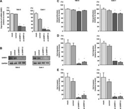
- Figure 5 Effects of UHRF1 knockdown in RCC cells and impact of UHRF1 expression on clinical RCC specimens ( A ) UHRF1 mRNA expression was determined at 72 h after transfection with si-UHRF1 . GUSB was used as an internal control. ( B ) UHRF1 protein expression was evaluated by western blotting at 72 h after transfection with si-UHRF1 . GAPDH was used as a loading control. * P < 0.0001. The bars indicate SDs. ( C ) Cell proliferation was assessed 72 h after transfection with si-UHRF1 using XTT assays. ( D ) Cell migration was assessed 48 h after transfection with si-UHRF1 using uncoated Transwell polycarbonate membrane filters. ( E ) Cell invasion was assessed 48 h after transfection with si-UHRF1 using Matrigel invasion assays. * P < 0.0001. The bars indicate SDs.
