 Explore
 Validate
 Learn
Learn Western blot
 Immunocytochemistry
Antibody data
- Antibody Data
- Antigen structure
- References [2]
- Comments [0]
- Validations
- Immunocytochemistry [1]
- Immunohistochemistry [1]
Submit
Validation data
Reference
Comment
Report error
- Product number
- HPA001466 - Provider product page

- Provider
- Atlas Antibodies
- Proper citation
- Atlas Antibodies Cat#HPA001466, RRID:AB_1855930
- Product name
- Anti-PTPN6
- Antibody type
- Polyclonal
- Description
- Polyclonal Antibody against Human PTPN6, Gene description: protein tyrosine phosphatase, non-receptor type 6, Alternative Gene Names: HCP, HCPH, PTP-1C, SHP-1, SHP1, Validated applications: WB, IHC, ICC, Uniprot ID: P29350, Storage: Store at +4°C for short term storage. Long time storage is recommended at -20°C.
- Reactivity
- Human, Rat
- Host
- Rabbit
- Conjugate
- Unconjugated
- Isotype
- IgG
- Vial size
- 100 µl
- Concentration
- 0.1 mg/ml
- Storage
- Store at +4°C for short term storage. Long time storage is recommended at -20°C.
- Handling
- The antibody solution should be gently mixed before use.
Submitted references Differential regulation of the transcriptomic and secretomic landscape of sensor and effector functions of human airway epithelial cells
Constitutive activation of STAT3 in Sézary syndrome is independent of SHP-1
Lehmann R, Müller M, Klassert T, Driesch D, Stock M, Heinrich A, Conrad T, Moore C, Schier U, Guthke R, Slevogt H
Mucosal Immunology 2018;11(3):627-642
Mucosal Immunology 2018;11(3):627-642
Constitutive activation of STAT3 in Sézary syndrome is independent of SHP-1
McKenzie R, Jones C, Tosi I, Caesar J, Whittaker S, Mitchell T
Leukemia 2011;26(2):323-331
Leukemia 2011;26(2):323-331
No comments: Submit comment
Supportive validation
- Submitted by
- Atlas Antibodies (provider)
- Main image
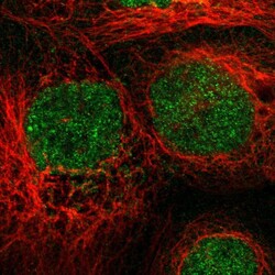
- Experimental details
- Immunofluorescent staining of human cell line A-431 shows localization to nucleus.
- Sample type
- Human

Supportive validation
- Submitted by
- Atlas Antibodies (provider)
- Enhanced method
- Orthogonal validation
- Main image
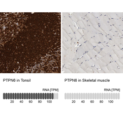
- Experimental details
- Immunohistochemistry analysis in human tonsil and skeletal muscle tissues using HPA001466 antibody. Corresponding PTPN6 RNA-seq data are presented for the same tissues.
- Sample type
- Human
- Protocol
- Protocol
