 Explore
 Validate
 Learn
Learn Immunocytochemistry
Antibody data
- Antibody Data
- Antigen structure
- References [2]
- Comments [0]
- Validations
- Immunocytochemistry [1]
Submit
Validation data
Reference
Comment
Report error
- Product number
- MAB1878 - Provider product page

- Provider
- R&D Systems
- Product name
- Human SHP-1 Antibody
- Antibody type
- Monoclonal
- Description
- Protein A or G purified from hybridoma culture supernatant. Detects human SHP-1.
- Reactivity
- Human
- Host
- Rat
- Conjugate
- Unconjugated
- Antigen sequence
P29350- Isotype
- IgG
- Antibody clone number
- 255402
- Vial size
- 100 ug
- Concentration
- LYOPH
- Storage
- Use a manual defrost freezer and avoid repeated freeze-thaw cycles. 12 months from date of receipt, -20 to -70 °C as supplied. 1 month, 2 to 8 °C under sterile conditions after reconstitution. 6 months, -20 to -70 °C under sterile conditions after reconstitution.
Submitted references The oncoprotein gankyrin promotes the development of colitis-associated cancer through activation of STAT3.
Regulation and function of immunosuppressive molecule human leukocyte antigen G5 in human bone tissue.
Sakurai T, Higashitsuji H, Kashida H, Watanabe T, Komeda Y, Nagai T, Hagiwara S, Kitano M, Nishida N, Abe T, Kiyonari H, Itoh K, Fujita J, Kudo M
Oncotarget 2017 Apr 11;8(15):24762-24776
Oncotarget 2017 Apr 11;8(15):24762-24776
Regulation and function of immunosuppressive molecule human leukocyte antigen G5 in human bone tissue.
Deschaseaux F, Gaillard J, Langonné A, Chauveau C, Naji A, Bouacida A, Rosset P, Heymann D, De Pinieux G, Rouas-Freiss N, Sensébé L
FASEB journal : official publication of the Federation of American Societies for Experimental Biology 2013 Aug;27(8):2977-87
FASEB journal : official publication of the Federation of American Societies for Experimental Biology 2013 Aug;27(8):2977-87
No comments: Submit comment
Supportive validation
- Submitted by
- R&D Systems (provider)
- Main image
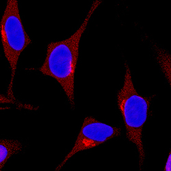
- Experimental details
- SHP-1 in HeLa Human Cell Line. SHP-1 was detected in immersion fixed HeLa human cervical epithelial carcinoma cell line using Rat Anti-Human SHP-1 Monoclonal Antibody (Catalog # MAB1878) at 3 µg/mL for 3 hours at room temperature. Cells were stained using the NorthernLights™ 557-conjugated Anti-Rat IgG Secondary Antibody (red; Catalog # NL013) and counterstained with DAPI (blue). Specific staining was localized to cytoplasm. View our protocol for Fluorescent ICC Staining of Cells on Coverslips.
