Explore
 Validate
 Learn
Learn Western blot
Antibody data
- Antibody Data
- Antigen structure
- References [3]
- Comments [0]
- Validations
- Western blot [3]
- Immunoprecipitation [1]
- Immunohistochemistry [2]
Submit
Validation data
Reference
Comment
Report error
- Product number
- NB100-60418 - Provider product page

- Provider
- Novus Biologicals
- Proper citation
- Novus Cat#NB100-60418, RRID:AB_905325
- Product name
- Rabbit Polyclonal CHD8 Antibody
- Antibody type
- Polyclonal
- Description
- Immunogen affinity purified.
- Reactivity
- Human, Mouse
- Host
- Rabbit
- Isotype
- IgG
- Vial size
- 0.1 ml
- Concentration
- 0.2 mg/ml
- Storage
- Store at 4C. Do not freeze.
Submitted references CHD8 regulates neurodevelopmental pathways associated with autism spectrum disorder in neural progenitors.
Frequent disruption of chromodomain helicase DNA-binding protein 8 (CHD8) and functionally associated chromatin regulators in prostate cancer.
Regulation of serum response factor activity and smooth muscle cell apoptosis by chromodomain helicase DNA-binding protein 8.
Sugathan A, Biagioli M, Golzio C, Erdin S, Blumenthal I, Manavalan P, Ragavendran A, Brand H, Lucente D, Miles J, Sheridan SD, Stortchevoi A, Kellis M, Haggarty SJ, Katsanis N, Gusella JF, Talkowski ME
Proceedings of the National Academy of Sciences of the United States of America 2014 Oct 21;111(42):E4468-77
Proceedings of the National Academy of Sciences of the United States of America 2014 Oct 21;111(42):E4468-77
Frequent disruption of chromodomain helicase DNA-binding protein 8 (CHD8) and functionally associated chromatin regulators in prostate cancer.
Damaschke NA, Yang B, Blute ML Jr, Lin CP, Huang W, Jarrard DF
Neoplasia (New York, N.Y.) 2014 Dec;16(12):1018-27
Neoplasia (New York, N.Y.) 2014 Dec;16(12):1018-27
Regulation of serum response factor activity and smooth muscle cell apoptosis by chromodomain helicase DNA-binding protein 8.
Rodenberg JM, Hoggatt AM, Chen M, Touw K, Jones R, Herring BP
American journal of physiology. Cell physiology 2010 Nov;299(5):C1058-67
American journal of physiology. Cell physiology 2010 Nov;299(5):C1058-67
No comments: Submit comment
Supportive validation
- Submitted by
- Novus Biologicals (provider)
- Main image

- Experimental details
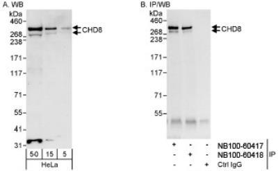
- Western Blot: CHD8 Antibody [NB100-60418] - Whole cell lysate (5, 15 and 50 ug for WB; 1 mg for IP, 20% of IP loaded) from HeLa cells. NB100-60418 used for WB at 0.04 ug/ml (A) and 1 ug/ml (B) and used for IP at 3 ug/mg lysate. CHD8 was also immunoprecipitated byNB100-60417, which recognizes an upstream epitope.
- Submitted by
- Novus Biologicals (provider)
- Main image
- Experimental details
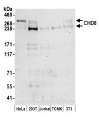
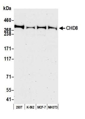
- Western Blot: CHD8 Antibody [NB100-60418] - Detection of Human and Mouse CHD8 by Western Blot. Samples: Whole cell lysate (50 ug) prepared using NETN buffer from HeLa, 293T, Jurkat, mouse TCMK-1, and mouse NIH3T3 cells. Antibodies: Affinity purified rabbit anti-CHD8 antibody NB100-60418 used for WB at 0.1 ug/ml. Detection: Chemiluminescence with an exposure time of 3 minutes.
- Submitted by
- Novus Biologicals (provider)
- Main image
- Experimental details
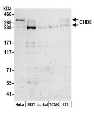
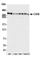
- Western Blot: CHD8 Antibody [NB100-60418] - Detection of human and mouse CHD8 by western blot. Samples: Whole cell lysate (50 ug) from HEK293T, K-562, MCF-7, and NIH 3T3 cells prepared using NETN lysis buffer. Antibody: Affinity purified Rabbit anti-CHD8 antibody NB100-60418 used for WB at 0.1 ug/ml. Detection: Chemiluminescence with an exposure time of 75 seconds.
Supportive validation
- Submitted by
- Novus Biologicals (provider)
- Main image
- Experimental details
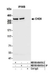
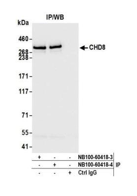
- Immunoprecipitation: CHD8 Antibody [NB100-60418] - Detection of human CHD8 by western blot of immunoprecipitates. Samples: Whole cell lysate (1.0 mg per IP reaction; 20% of IP loaded) from HEK293T cells prepared using NETN lysis buffer. Antibodies: Rabbit anti-CHD8 antibody NB100-60418 (lot NB100-60418-4) used for IP at 6 ul per reaction. CHD8 was also immunoprecipitated by a previous lot of this antibody (lot NB100-60418-3). For blotting immunoprecipitated CHD8, NB100-60418 was used at 0.1 ug/ml. Chemiluminescence with an exposure time of 1 second.
Supportive validation
- Submitted by
- Novus Biologicals (provider)
- Main image
- Experimental details
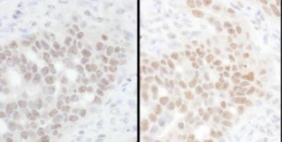
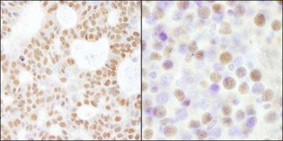
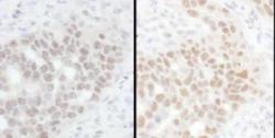
- Immunohistochemistry-Paraffin: CHD8 Antibody [NB100-60418] - Human skin basal cell carcinoma (left) and mouse renal cell carcinoma (right). Antibody used at a dilution of 1:200 (1ug/ml).
- Submitted by
- Novus Biologicals (provider)
- Main image
- Experimental details
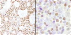
- Immunohistochemistry-Paraffin: CHD8 Antibody [NB100-60418] - Human ovarian carcinoma. Antibody: Affinity purified rabbit anti-CHD8 used at a dilution of 1:200 (1ug/ml). Detection: DAB