 Explore
 Validate
 Learn
Learn Flow cytometry
Antibody data
- Antibody Data
- Antigen structure
- References [3]
- Comments [0]
- Validations
- Flow cytometry [1]
Submit
Validation data
Reference
Comment
Report error
- Product number
- 50-9076-42 - Provider product page

- Provider
- Invitrogen Antibodies
- Product name
- Phospho-LCK (Tyr505) Monoclonal Antibody (SRRCHA), eFluor™ 660, eBioscience™
- Antibody type
- Monoclonal
- Antigen
- Synthetic peptide
- Description
- Description: This SRRCHA monoclonal antibody recognizes human and mouse LCK when phosphorylated on tyrosine 505 (Y505). Y505 is a negative-regulatory site that is phosphorylated by the kinase CSK. LCK is expressed in T cells where it associates with the cytoplasmic tails of the CD4 and CD8 co-receptors on T helper cells and cytotoxic T cells, respectively. When the TCR is engaged, LCK acts to phosphorylate the intracellular chains of CD3 and zeta chains of the TCR complex, allowing the cytoplasmic tyrosine kinase, ZAP-70, to bind to them. LCK then phosphorylates and activates ZAP-70, which then phosphorylates the adaptor protein, LAT, that serves as a docking site for a number of other proteins, including SLP-76, Grb2, and phospholipase C gamma1. Mice lacking expression of LCK show defects in T cell development and function. Applications Reported:This SRRCHA antibody has been reported for use in intracellular staining followed by flow cytometric analysis. Applications Tested: This SRRCHA antibody has been pre-titrated and tested by intracellular staining followed by flow cytometric analysis of stimulated normal human peripheral blood cells. This can be used at 5 µL (0.125 µg) per test. A test is defined as the amount (µg) of antibody that will stain a cell sample in a final volume of 100 µL. Cell number should be determined empirically but can range from 10^5 to 10^8 cells/test. eFluor® 660 is a replacement for Alexa Fluor® 647. eFluor® 660 emits at 659 nm and is excited with the red laser (633 nm). Please make sure that your instrument is capable of detecting this fluorochrome. Staining Protocol: All protocols work well for this monoclonal antibody. Use of Protocol A: Two-step protocol: intracellular (cytoplasmic) proteins allows for the greatest flexibility for detection of surface and intracellular (cytoplasmic) proteins. Use of Protocol B: One-step protocol: intracellular (nuclear) proteins is recommended for staining of transcription factors in conjunction with surface and phosphorylated intracellular (cytoplasmic) proteins. Protocol C: Two-step protocol: Fixation/Methanol allows for the greatest discrimination of phospho-specific signaling between unstimulated and stimulated samples, but with limitations on the ability to stain specific surface proteins (refer to "Clone Performance Following Fixation/Permeabilization" located in the BestProtocols Section under the Resources tab online). All Protocols can be found in the Flow Cytometry Protocols: "Staining Intracellular Antigens for Flow Cytometry Protocol" located in the BestProtocols® Section under the Resources tab online. Excitation: 633-647 nm; Emission: 668 nm; Laser: Red Laser. Filtration: 0.2 µm post-manufacturing filtered.
- Reactivity
- Human, Mouse
- Host
- Mouse
- Isotype
- IgG
- Antibody clone number
- SRRCHA
- Vial size
- 100 Tests
- Concentration
- 5 μL/Test
- Storage
- 4°C, store in dark, DO NOT FREEZE!
Submitted references Feedback circuits monitor and adjust basal Lck-dependent events in T cell receptor signaling.
Function of the Src-family kinases, Lck and Fyn, in T-cell development and activation.
Regulation of Lck activity by CD4 and CD28 in the immunological synapse.
Schoenborn JR, Tan YX, Zhang C, Shokat KM, Weiss A
Science signaling 2011 Sep 13;4(190):ra59
Science signaling 2011 Sep 13;4(190):ra59
Function of the Src-family kinases, Lck and Fyn, in T-cell development and activation.
Palacios EH, Weiss A
Oncogene 2004 Oct 18;23(48):7990-8000
Oncogene 2004 Oct 18;23(48):7990-8000
Regulation of Lck activity by CD4 and CD28 in the immunological synapse.
Holdorf AD, Lee KH, Burack WR, Allen PM, Shaw AS
Nature immunology 2002 Mar;3(3):259-64
Nature immunology 2002 Mar;3(3):259-64
No comments: Submit comment
Supportive validation
- Submitted by
- Invitrogen Antibodies (provider)
- Main image
- Experimental details
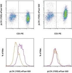
- TOP: Intracellular staining of untreated (left) or 5-minute hydrogen peroxide-activated sodium pervanadate-treated (right) normal human peripheral blood cells with Atni-Human CD3 PE (Product # 12-0037-42) and Anti-Human/Mouse phospho-LCK (Y505) eFluor® 660, using the Intracellular Fixation & Permeabilization Buffer Set (Product # 88-8824-00) and protocol. Cells in the lymphocytes gate were used for analysis. BOTTOM: Intracellular staining of untreated (orange histogram) or 5-minute hydrogen peroxide-activated sodium pervanadate-treated (purple histogram) normal human peripheral blood cells with Anti-Human CD3 PE (Product # 12-0037-42), Anti-Human CD19 FITC (Product # 11-0199-42), and Anti-Human/Mouse phospho-LCK (Y505) eFluor® 660, using the Intracellular Fixation & Permeabilization Buffer Set (Product # 88-8824-00) and protocol. Lymphocytes in the CD3+ (left) or CD19+ (right) gates were used for analysis.
