Explore
 Validate
 Learn
Learn Western blot
Antibody data
- Antibody Data
- Antigen structure
- References [0]
- Comments [0]
- Validations
- Western blot [3]
Submit
Validation data
Reference
Comment
Report error
- Product number
- MA1-41203 - Provider product page

- Provider
- Invitrogen Antibodies
- Product name
- LCK Monoclonal Antibody (33D196)
- Antibody type
- Monoclonal
- Antigen
- Synthetic peptide
- Description
- The immunogen sequence is 100% homologus in mouse. Suggested positive control: Jurkat whole cell lysate.
- Reactivity
- Human, Mouse
- Host
- Mouse
- Isotype
- IgG
- Antibody clone number
- 33D196
- Vial size
- 200 μL
- Concentration
- 1.0 mg/mL
- Storage
- Store at 4°C short term. For long term storage, store at -20°C, avoiding freeze/thaw cycles.
No comments: Submit comment
Supportive validation
- Submitted by
- Invitrogen Antibodies (provider)
- Main image
- Experimental details
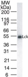
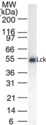
- Western blot analysis of LCK in 20 µg/lane Jurkat cell lysate. Samples were incubated in LCK monoclonal antibody (Product # MA1-41203) using a dilution of 1:1000.
- Submitted by
- Invitrogen Antibodies (provider)
- Main image
- Experimental details
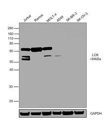
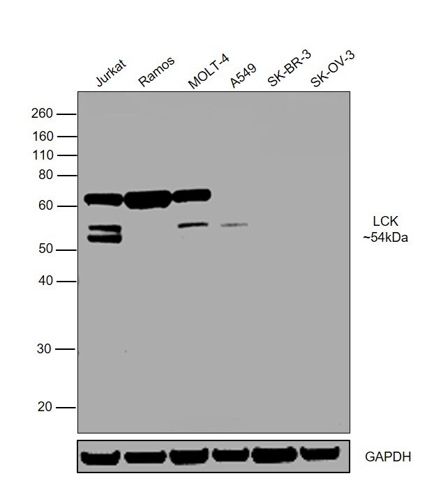
- Western blot was performed using Anti-LCK Monoclonal Antibody (33D196) (Product # MA1-41203) and a 54kDa band corresponding to LCK was observed in Jurkat, MOLT-4 and A549, but not in Ramos, SK-BR-3 and SK-OV-3. An uncharacterized band was alos observed at ~ 65kDa in few cell lines. Whole cell lysates (30ug lysate) of Jurkat (Lane 1), Ramos (Lane 2), MOLT-4 (Lane 3), A549 (Lane 4), SK-BR-3 (Lane 5) and SK-OV-3 (Lane 6) were electrophoresed using Novex® NuPAGE® 4-12 % Bis-Tris gel (Product # NP0322BOX). Resolved proteins were then transferred onto a nitrocellulose membrane (Product # IB23001) by iBlot® 2 Dry Blotting System (Product # IB21001). The bot was probed with the primary antibody (1:1000 dilution) detected by chemiluminescence with Goat anti-Mouse IgG (H+L) Superclonal™ Recombinant Secondary Antibody, HRP (Product # A28177, 1:4000 dilution) using the iBright FL 1000 (Product # A32752). Chemiluminescent detection was performed using Novex® ECL Chemiluminescent Substrate Reagent Kit (Product # WP20005).
- Submitted by
- Invitrogen Antibodies (provider)
- Main image

- Experimental details
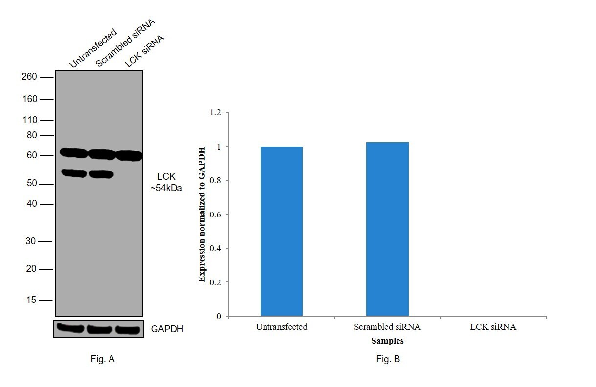
- Knockdown of LCK was achieved by transfecting Jurkat with LCK specific siRNAs (Silencer® select Products # s8108, s8107). Western blot analysis (Fig. a) was performed using whole cell extracts from the LCK knockdown cells (Lane 3), non-specific scrambled siRNA transfected cells (Lane 2) and untransfected cells (Lane 1). The blot was probed with LCK Monoclonal Antibody (33D196) (Product # MA1-41203, 1:1000 dilution) and Goat anti-Mouse IgG (H+L) Superclonal™ Recombinant Secondary Antibody, HRP (Product # A28177, 1:4000 dilution). Densitometric analysis of this western blot is shown in histogram (Fig. b). Decrease in signal upon siRNA mediated knock down confirms that antibody is specific to LCK. An uncharacterized band was observed at ~60kDa.