 Explore
 Validate
 Learn
Learn Western blot
 Immunohistochemistry
Antibody data
- Antibody Data
- Antigen structure
- References [7]
- Comments [0]
- Validations
- Immunohistochemistry [1]
- Flow cytometry [2]
- Other assay [6]
Submit
Validation data
Reference
Comment
Report error
- Product number
- MA5-17687 - Provider product page

- Provider
- Invitrogen Antibodies
- Product name
- CD45 Monoclonal Antibody (YAML501.4)
- Antibody type
- Monoclonal
- Description
- MA5-17687 targets CD45/PTPRC in FACS and WB applications and shows reactivity with Human samples. The MA5-17687 immunogen is rat monoclonal antibody raised against acute myeloid leukemia cells of human origin.
- Reactivity
- Human
- Host
- Rat
- Isotype
- IgG
- Antibody clone number
- YAML501.4
- Vial size
- 200 µg
- Concentration
- 1 mg/mL
- Storage
- Store at 4°C short term. For long term storage, store at -20°C, avoiding freeze/thaw cycles.
Submitted references Versican promotes T helper 17 cytotoxic inflammation and impedes oligodendrocyte precursor cell remyelination.
Expression of antioxidant enzymes in lesions of multiple sclerosis and its models.
Integration of Functional Imaging, Cytometry, and Unbiased Proteomics Reveals New Features of Endothelial-to-Mesenchymal Transition in Ischemic Mitral Valve Regurgitation in Human Patients.
Chronic mTOR activation induces a degradative smooth muscle cell phenotype.
Evolution of Estrogen Receptor Status from Primary Tumors to Metastasis and Serially Collected Circulating Tumor Cells.
The tyrosine phosphatase SHP2 controls TGFβ-induced STAT3 signaling to regulate fibroblast activation and fibrosis.
Filtration-based enrichment of circulating tumor cells from all prostate cancer risk groups.
Ghorbani S, Jelinek E, Jain R, Buehner B, Li C, Lozinski BM, Sarkar S, Kaushik DK, Dong Y, Wight TN, Karimi-Abdolrezaee S, Schenk GJ, Strijbis EM, Geurts J, Zhang P, Ling CC, Yong VW
Nature communications 2022 May 4;13(1):2445
Nature communications 2022 May 4;13(1):2445
Expression of antioxidant enzymes in lesions of multiple sclerosis and its models.
Moezzi D, Dong Y, Jain RW, Lozinski BM, Ghorbani S, D'Mello C, Wee Yong V
Scientific reports 2022 Jul 26;12(1):12761
Scientific reports 2022 Jul 26;12(1):12761
Integration of Functional Imaging, Cytometry, and Unbiased Proteomics Reveals New Features of Endothelial-to-Mesenchymal Transition in Ischemic Mitral Valve Regurgitation in Human Patients.
Lupieri A, Nagata Y, Passos LSA, Beker-Greene D, Kirkwood KA, Wylie-Sears J, Alvandi Z, Higashi H, Hung JW, Singh SA, Bischoff J, Levine RA, Aikawa E
Frontiers in cardiovascular medicine 2021;8:688396
Frontiers in cardiovascular medicine 2021;8:688396
Chronic mTOR activation induces a degradative smooth muscle cell phenotype.
Li G, Wang M, Caulk AW, Cilfone NA, Gujja S, Qin L, Chen PY, Chen Z, Yousef S, Jiao Y, He C, Jiang B, Korneva A, Bersi MR, Wang G, Liu X, Mehta S, Geirsson A, Gulcher JR, Chittenden TW, Simons M, Humphrey JD, Tellides G
The Journal of clinical investigation 2020 Mar 2;130(3):1233-1251
The Journal of clinical investigation 2020 Mar 2;130(3):1233-1251
Evolution of Estrogen Receptor Status from Primary Tumors to Metastasis and Serially Collected Circulating Tumor Cells.
Forsare C, Bendahl PO, Moberg E, Levin Tykjær Jørgensen C, Jansson S, Larsson AM, Aaltonen K, Rydén L
International journal of molecular sciences 2020 Apr 20;21(8)
International journal of molecular sciences 2020 Apr 20;21(8)
The tyrosine phosphatase SHP2 controls TGFβ-induced STAT3 signaling to regulate fibroblast activation and fibrosis.
Zehender A, Huang J, Györfi AH, Matei AE, Trinh-Minh T, Xu X, Li YN, Chen CW, Lin J, Dees C, Beyer C, Gelse K, Zhang ZY, Bergmann C, Ramming A, Birchmeier W, Distler O, Schett G, Distler JHW
Nature communications 2018 Aug 14;9(1):3259
Nature communications 2018 Aug 14;9(1):3259
Filtration-based enrichment of circulating tumor cells from all prostate cancer risk groups.
Awe JA, Saranchuk J, Drachenberg D, Mai S
Urologic oncology 2017 May;35(5):300-309
Urologic oncology 2017 May;35(5):300-309
No comments: Submit comment
Supportive validation
- Submitted by
- Invitrogen Antibodies (provider)
- Main image

- Experimental details
- Immunohistochemical staining of paraformaldehyde-fixed paraffin-embedded human spleen sections at 400x magnification with DAPI counterstain. Primary antibody used was a CD45/PTPRC monoclonal antibody (clone YAML501.4) at a 1:500 dilution, followed by secondary antibody was a 488 fluorescent conjugated Goat anti-rat at a 1:500 dilution and Normal Goat Serum (blocking)

Supportive validation
- Submitted by
- Invitrogen Antibodies (provider)
- Main image

- Experimental details
- Flow cytometric analysis of human peripheral blood lymphocytes were stained using a CD45/PTPRC monoclonal antibody (Product # MA5-17687) (filled histogram) or rat IgG2a isotype control (open histogram).

- Submitted by
- Invitrogen Antibodies (provider)
- Main image
- Experimental details
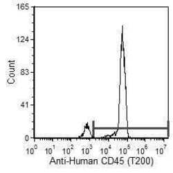
- Flow cytometric analysis of Human peripheral blood leukocytes staining using anti-CD45/PTPRC Monoclonal Antibody (Product # MA5-17687) at a 0.5 µg/10^6 cells dilution.

Supportive validation
- Submitted by
- Invitrogen Antibodies (provider)
- Main image

- Experimental details
- NULL

- Submitted by
- Invitrogen Antibodies (provider)
- Main image

- Experimental details
- Fig. 1 Decreased expression of SHP2 in SSc. a The mRNA levels of SHP2 are significantly reduced in SSc skin as compared to healthy skin ( n = 7). b Immunohistochemistry of SHP2 in SSc skin and matched healthy controls. Representative images are shown at 200- and 1000-fold magnification. c Immunofluorescence staining of SHP2 with co-staining for the fibroblast marker P4Hbeta, the endothelial cell marker CD31 and the leukocyte marker CD45, and DAPI. SSc fibroblasts demonstrated a reduced staining for SHP2 compared to healthy control. Representative images are shown at 400-fold magnification. Immunofluorescence pictures were processed to generate Voronoi tessellated pictures amenable to computational simulation. Quantification of SHP2 staining intensity ( n = 5) and of SHP2-positive cells ( n = 5). d , e The mRNA ( n = 5) ( d ) and protein level ( n = 4) ( e ) of SHP2 are decreased in cultured SSc fibroblasts. Horizontal scale bar, for all images, 500 mum. All data are presented as median +- s.e.m. The p values are expressed as follows: 0.05 > p > 0.01*; 0.01 > p > 0.001**; p < 0.001***. Significance was determined by Mann-Whitney test. SSc: systemic sclerosis, Healthy: healthy individual, int.: intensity

- Submitted by
- Invitrogen Antibodies (provider)
- Main image
- Experimental details
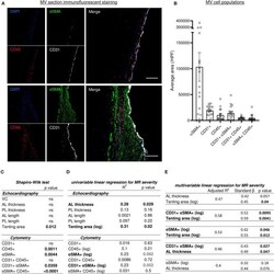
- Figure 4 Histo-cytometry assessment of MV cell composition correlated with echocardiography measures revealed that MR is concomitantly associated to EndMT and leaflet's thickness or tenting. (A) Representative MV section from patient with different range of cellular composition, (patients #14 upper and #12 bottom) stained with immunofluorescent co-staining of DAPI (blue), alphaSMA (green), CD45 (red), and CD31 (white), merge on the left panel (scale bar represents 100 mum). (B) Cell count per high power field (HPF) for alphaSMA single positive cells (alphaSMA+), CD45 single positive cells (CD45+), CD31 single positive cells (CD31+), along with CD31/alphaSMA double positive (CD31+ alphaSMA+) and CD31/CD45 double positive (CD31+ CD45+). (C) Shapiro-Wilk test of data distribution. (D) Univariable linear regression with VC. (E) Multivariable linear regression with VC. MV, mitral valves; VC, vena contracta; PL, posterior mitral leaflet; AL, anterior mitral leaflet; SP, single positive.

- Submitted by
- Invitrogen Antibodies (provider)
- Main image
- Experimental details
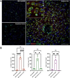
- Expression of SOD1 antioxidant enzyme is elevated in the chronic active rim of MS lesions. ( A ) Representative confocal images of NAWM, inactive Core, and active rim of an MS lesion labeled with CD45 for immune cells (green), SOD1 (red) and DAPI (blue). ( B ) Bar graphs comparing the percent of field of view (FOV) that is CD45 + , SOD1 + , and CD45 + SOD1 + . Scale bar = 100 um. Data are shown as mean +- S.D, n = 3 MS patients. Significance indicated as * p < 0.05, ** p < 0.01, *** p < 0.001, **** p < 0.0001, One-way ANOVA with Tukey's post-hoc test.

- Submitted by
- Invitrogen Antibodies (provider)
- Main image
- Experimental details
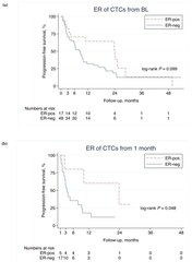
- Figure 4 ER-status on CTCs and PFS. Association of progression-free survival (PFS) and the estrogen receptor (ER) status at different timepoints using the circulating tumor cell (CTC) Drop-Mount method. Note that the time-axes start at 0, 1, and 3 months, respectively, in ( a ), ( b ), and ( c ) (landmark analysis). * p -values are from the log-rank test.

- Submitted by
- Invitrogen Antibodies (provider)
- Main image

- Experimental details
- Figure 5 Immunofluorescent staining of CTCs in metastatic breast cancer blood samples. Circulating tumor cells (CTCs) were scanned using ( a ) BX63 Upright Microscope; ( a ) a composite image of all channels, ( b ) 4',6-diamidino-2-phenylindole (DAPI) counterstain (fluorescent blue), ( c ) estrogen receptor alpha stained with AlexaFluor488 (green), ( d ) cytokeratins 8, 18, and 19 stained with phycoerythrin (red), and ( e ) CD45 stained with AlexaFluor647 (yellow).
