 Explore
 Validate
 Learn
Learn Flow cytometry
Antibody data
- Antibody Data
- Antigen structure
- References [8]
- Comments [0]
- Validations
- Flow cytometry [1]
- Other assay [2]
Submit
Validation data
Reference
Comment
Report error
- Product number
- MHCD4512 - Provider product page

- Provider
- Invitrogen Antibodies
- Product name
- CD45 Monoclonal Antibody (HI30), PE-Cyanine7
- Antibody type
- Monoclonal
- Antigen
- Other
- Description
- The R-phycoerythrin (PE)-Cy7® tandem conjugate permits simultaneous multicolor labeling and detection of multiple targets with excitation by a 488 nm laser without occupying traditional channels associated with FITC and PE. Alternatively, the PE dye tandem can be excited using a 532 nm or 561 nm laser.
- Reactivity
- Human
- Host
- Mouse
- Isotype
- IgG
- Antibody clone number
- HI30
- Vial size
- 500 µL
- Storage
- 4° C, store in dark
Submitted references Transcriptional, epigenetic and metabolic signatures in cardiometabolic syndrome defined by extreme phenotypes.
Mesenchymal stem cells negatively regulate CD4(+) T cell activation in patients with primary Sjögren syndrome through the miRNA‑125b and miRNA‑155 TCR pathway.
miR-9 Does Not Regulate Lamin A Expression in Metastatic Cells from Lung Adenocarcinoma.
Elucidating the fundamental fibrotic processes driving abdominal adhesion formation.
Low lamin A expression in lung adenocarcinoma cells from pleural effusions is a pejorative factor associated with high number of metastatic sites and poor Performance status.
Methylation Analysis in Distinct Immune Cell Subsets in Type 1 Diabetes.
Increased DNA methylation variability in type 1 diabetes across three immune effector cell types.
Dynamic change in natural killer cell type in the human ocular mucosa in situ as means of immune evasion by adenovirus infection.
Seyres D, Cabassi A, Lambourne JJ, Burden F, Farrow S, McKinney H, Batista J, Kempster C, Pietzner M, Slingsby O, Cao TH, Quinn PA, Stefanucci L, Sims MC, Rehnstrom K, Adams CL, Frary A, Ergüener B, Kreuzhuber R, Mocciaro G, D'Amore S, Koulman A, Grassi L, Griffin JL, Ng LL, Park A, Savage DB, Langenberg C, Bock C, Downes K, Wareham NJ, Allison M, Vacca M, Kirk PDW, Frontini M
Clinical epigenetics 2022 Mar 12;14(1):39
Clinical epigenetics 2022 Mar 12;14(1):39
Mesenchymal stem cells negatively regulate CD4(+) T cell activation in patients with primary Sjögren syndrome through the miRNA‑125b and miRNA‑155 TCR pathway.
Gong B, Zheng L, Lu Z, Huang J, Pu J, Pan S, Zhang M, Liu J, Tang J
Molecular medicine reports 2021 Jan;23(1)
Molecular medicine reports 2021 Jan;23(1)
miR-9 Does Not Regulate Lamin A Expression in Metastatic Cells from Lung Adenocarcinoma.
Guinde J, Benoit A, Frankel D, Robert S, Ostacolo K, Lévy N, Astoul P, Roll P, Kaspi E
International journal of molecular sciences 2020 Feb 26;21(5)
International journal of molecular sciences 2020 Feb 26;21(5)
Elucidating the fundamental fibrotic processes driving abdominal adhesion formation.
Foster DS, Marshall CD, Gulati GS, Chinta MS, Nguyen A, Salhotra A, Jones RE, Burcham A, Lerbs T, Cui L, King ME, Titan AL, Ransom RC, Manjunath A, Hu MS, Blackshear CP, Mascharak S, Moore AL, Norton JA, Kin CJ, Shelton AA, Januszyk M, Gurtner GC, Wernig G, Longaker MT
Nature communications 2020 Aug 13;11(1):4061
Nature communications 2020 Aug 13;11(1):4061
Low lamin A expression in lung adenocarcinoma cells from pleural effusions is a pejorative factor associated with high number of metastatic sites and poor Performance status.
Kaspi E, Frankel D, Guinde J, Perrin S, Laroumagne S, Robaglia-Schlupp A, Ostacolo K, Harhouri K, Tazi-Mezalek R, Micallef J, Dutau H, Tomasini P, De Sandre-Giovannoli A, Lévy N, Cau P, Astoul P, Roll P
PloS one 2017;12(8):e0183136
PloS one 2017;12(8):e0183136
Methylation Analysis in Distinct Immune Cell Subsets in Type 1 Diabetes.
Dang MN, Bradford CM, Pozzilli P, Leslie RD
Methods in molecular biology (Clifton, N.J.) 2016;1433:143-51
Methods in molecular biology (Clifton, N.J.) 2016;1433:143-51
Increased DNA methylation variability in type 1 diabetes across three immune effector cell types.
Paul DS, Teschendorff AE, Dang MA, Lowe R, Hawa MI, Ecker S, Beyan H, Cunningham S, Fouts AR, Ramelius A, Burden F, Farrow S, Rowlston S, Rehnstrom K, Frontini M, Downes K, Busche S, Cheung WA, Ge B, Simon MM, Bujold D, Kwan T, Bourque G, Datta A, Lowy E, Clarke L, Flicek P, Libertini E, Heath S, Gut M, Gut IG, Ouwehand WH, Pastinen T, Soranzo N, Hofer SE, Karges B, Meissner T, Boehm BO, Cilio C, Elding Larsson H, Lernmark Å, Steck AK, Rakyan VK, Beck S, Leslie RD
Nature communications 2016 Nov 29;7:13555
Nature communications 2016 Nov 29;7:13555
Dynamic change in natural killer cell type in the human ocular mucosa in situ as means of immune evasion by adenovirus infection.
Yawata N, Selva KJ, Liu YC, Tan KP, Lee AW, Siak J, Lan W, Vania M, Arundhati A, Tong L, Li J, Mehta JS, Yawata M
Mucosal immunology 2016 Jan;9(1):159-70
Mucosal immunology 2016 Jan;9(1):159-70
No comments: Submit comment
Supportive validation
- Submitted by
- Invitrogen Antibodies (provider)
- Main image
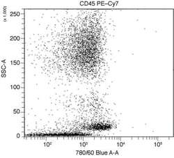
- Experimental details
- Ammonium chloride lysed whole blood was stained with mouse anti human CD45 PE-Cy7 (Product # MHCD4512), washed, and analyzed on a flow cytometer with a 488nm excitation source.

Supportive validation
- Submitted by
- Invitrogen Antibodies (provider)
- Main image

- Experimental details
- Fig 3 Flow cytometry analysis of lamin A and EMA in adenocarcinoma cells. (A to E) Representative results of lamin A expression in malignant cells contained in 2 metastatic pleural effusions from lung adenocarcinoma (left panel = Patient (Pt) 16 and right panel = Pt 45) using flow cytometry. Positivity thresholds were defined using isotype controls. Cell analysis strategy: (A) Cells were selected using SSC and FSC criteria. (B) Live cells lacking CD45 expression were considered adenocarcinoma cells (CD45-/EMA- and CD45-/EMA+ cells). Leukocytes (CD45+/EMA- cells) were excluded from analysis. (C and D) Lamin A and EMA expression in adenocarcinoma cells. See also Table 2 .(E) Correlation of EMA and lamin A expression in adenocarcinoma cells: The percentage of lamin A-expressing adenocarcinoma cells was positively correlated with the percentage of EMA-expressing cells (p = 0.0123; Spearman test).

- Submitted by
- Invitrogen Antibodies (provider)
- Main image

- Experimental details
- Figure 2 Flow cytometry analysis of lamin A and epithelial membrane antigen (EMA) in adenocarcinoma cells from pleural effusions. Representative results of lamin A and EMA expression in malignant cells contained in metastatic pleural effusions from lung adenocarcinoma (left panels = Patients (Pt) 59 and 65, from the ''Low Lamin A expression'' group and right panels = Pt 60, 70, and 71, from the ''High Lamin A expression'' group) using flow cytometry. Positivity thresholds were defined using isotype controls.
