Explore
 Validate
 Learn
Learn Flow cytometry
 Other assay
Antibody data
- Antibody Data
- Antigen structure
- References [12]
- Comments [0]
- Validations
- Other assay [11]
Submit
Validation data
Reference
Comment
Report error
- Product number
- MHCD4520 - Provider product page

- Provider
- Invitrogen Antibodies
- Product name
- CD45 Monoclonal Antibody (HI30), Alexa Fluor™ 488
- Antibody type
- Monoclonal
- Antigen
- Other
- Description
- Based on our testing, publications, and results reported from customers, the Alexa Fluor® 488 dye provides the best fluorescein (FITC) substitute.
- Reactivity
- Human
- Host
- Mouse
- Conjugate
- Green dye
- Isotype
- IgG
- Antibody clone number
- HI30
- Vial size
- 500 µL
- Storage
- 4° C, store in dark
Submitted references Tracking the expression of therapeutic protein targets in rare cells by antibody-mediated nanoparticle labelling and magnetic sorting.
ALICE: a hybrid AI paradigm with enhanced connectivity and cybersecurity for a serendipitous encounter with circulating hybrid cells.
Transient non-integrative expression of nuclear reprogramming factors promotes multifaceted amelioration of aging in human cells.
Mitochondrial Transfer of Wharton's Jelly Mesenchymal Stem Cells Eliminates Mutation Burden and Rescues Mitochondrial Bioenergetics in Rotenone-Stressed MELAS Fibroblasts.
CTC phenotyping for a preoperative assessment of tumor metastasis and overall survival of pancreatic ductal adenocarcinoma patients.
A patient derived xenograft model of cervical cancer and cervical dysplasia.
Single-cell mRNA cytometry via sequence-specific nanoparticle clustering and trapping.
Bioengineered constructs combined with exercise enhance stem cell-mediated treatment of volumetric muscle loss.
Deformability-based cell selection with downstream immunofluorescence analysis.
An artificial niche preserves the quiescence of muscle stem cells and enhances their therapeutic efficacy.
Dynamic change in natural killer cell type in the human ocular mucosa in situ as means of immune evasion by adenovirus infection.
Deformability of Tumor Cells versus Blood Cells.
Labib M, Wang Z, Ahmed SU, Mohamadi RM, Duong B, Green B, Sargent EH, Kelley SO
Nature biomedical engineering 2021 Jan;5(1):41-52
Nature biomedical engineering 2021 Jan;5(1):41-52
ALICE: a hybrid AI paradigm with enhanced connectivity and cybersecurity for a serendipitous encounter with circulating hybrid cells.
Cheng KS, Pan R, Pan H, Li B, Meena SS, Xing H, Ng YJ, Qin K, Liao X, Kosgei BK, Wang Z, Han RPS
Theranostics 2020;10(24):11026-11048
Theranostics 2020;10(24):11026-11048
Transient non-integrative expression of nuclear reprogramming factors promotes multifaceted amelioration of aging in human cells.
Sarkar TJ, Quarta M, Mukherjee S, Colville A, Paine P, Doan L, Tran CM, Chu CR, Horvath S, Qi LS, Bhutani N, Rando TA, Sebastiano V
Nature communications 2020 Mar 24;11(1):1545
Nature communications 2020 Mar 24;11(1):1545
Mitochondrial Transfer of Wharton's Jelly Mesenchymal Stem Cells Eliminates Mutation Burden and Rescues Mitochondrial Bioenergetics in Rotenone-Stressed MELAS Fibroblasts.
Lin TK, Chen SD, Chuang YC, Lan MY, Chuang JH, Wang PW, Hsu TY, Wang FS, Tsai MH, Huang ST, Wang XW, Tsai PC, Lin HY, Liou CW
Oxidative medicine and cellular longevity 2019;2019:9537504
Oxidative medicine and cellular longevity 2019;2019:9537504
CTC phenotyping for a preoperative assessment of tumor metastasis and overall survival of pancreatic ductal adenocarcinoma patients.
Sun Y, Wu G, Cheng KS, Chen A, Neoh KH, Chen S, Tang Z, Lee PF, Dai M, Han RPS
EBioMedicine 2019 Aug;46:133-149
EBioMedicine 2019 Aug;46:133-149
A patient derived xenograft model of cervical cancer and cervical dysplasia.
Larmour LI, Cousins FL, Teague JA, Deane JA, Jobling TW, Gargett CE
PloS one 2018;13(10):e0206539
PloS one 2018;13(10):e0206539
Single-cell mRNA cytometry via sequence-specific nanoparticle clustering and trapping.
Labib M, Mohamadi RM, Poudineh M, Ahmed SU, Ivanov I, Huang CL, Moosavi M, Sargent EH, Kelley SO
Nature chemistry 2018 May;10(5):489-495
Nature chemistry 2018 May;10(5):489-495
Bioengineered constructs combined with exercise enhance stem cell-mediated treatment of volumetric muscle loss.
Quarta M, Cromie M, Chacon R, Blonigan J, Garcia V, Akimenko I, Hamer M, Paine P, Stok M, Shrager JB, Rando TA
Nature communications 2017 Jun 20;8:15613
Nature communications 2017 Jun 20;8:15613
Deformability-based cell selection with downstream immunofluorescence analysis.
Shaw Bagnall J, Byun S, Miyamoto DT, Kang JH, Maheswaran S, Stott SL, Toner M, Manalis SR
Integrative biology : quantitative biosciences from nano to macro 2016 May 16;8(5):654-64
Integrative biology : quantitative biosciences from nano to macro 2016 May 16;8(5):654-64
An artificial niche preserves the quiescence of muscle stem cells and enhances their therapeutic efficacy.
Quarta M, Brett JO, DiMarco R, De Morree A, Boutet SC, Chacon R, Gibbons MC, Garcia VA, Su J, Shrager JB, Heilshorn S, Rando TA
Nature biotechnology 2016 Jul;34(7):752-9
Nature biotechnology 2016 Jul;34(7):752-9
Dynamic change in natural killer cell type in the human ocular mucosa in situ as means of immune evasion by adenovirus infection.
Yawata N, Selva KJ, Liu YC, Tan KP, Lee AW, Siak J, Lan W, Vania M, Arundhati A, Tong L, Li J, Mehta JS, Yawata M
Mucosal immunology 2016 Jan;9(1):159-70
Mucosal immunology 2016 Jan;9(1):159-70
Deformability of Tumor Cells versus Blood Cells.
Shaw Bagnall J, Byun S, Begum S, Miyamoto DT, Hecht VC, Maheswaran S, Stott SL, Toner M, Hynes RO, Manalis SR
Scientific reports 2015 Dec 18;5:18542
Scientific reports 2015 Dec 18;5:18542
No comments: Submit comment
Supportive validation
- Submitted by
- Invitrogen Antibodies (provider)
- Main image
- Experimental details
- NULL
- Conjugate
- Green dye
- Submitted by
- Invitrogen Antibodies (provider)
- Main image

- Experimental details
- NULL
- Conjugate
- Green dye
- Submitted by
- Invitrogen Antibodies (provider)
- Main image
- Experimental details
- NULL
- Conjugate
- Green dye
- Submitted by
- Invitrogen Antibodies (provider)
- Main image
- Experimental details
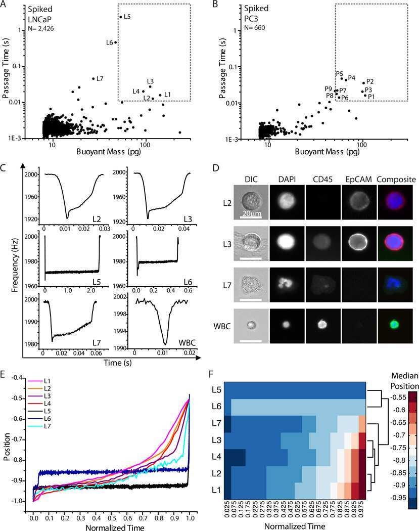
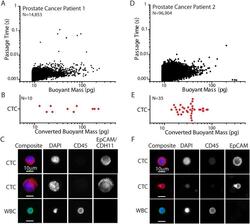
- Figure 5 Human patient CTC deformability. ( A ) SMR measurement of prostate cancer Patient 1 blood sample, after having been processed in the CTC-iChip 14 15 . ( B ) Based on fluorescence images, the diameter of each CTC was converted to an approximate buoyant mass value to visualize where they fall among the data measured by the SMR as shown in ( A ). Each point represents one cell. ( C ) A sampling of fluorescence images of CTCs and a white blood cell (WBC) taken from the same prostate cancer patient sample after it was measured in the SMR. ( D ) SMR measurement of prostate cancer Patient 2 blood sample, after having been processed in the CTC-iChip. ( E ) Based on fluorescence images, the diameter of each CTC was converted to an approximate buoyant mass value. Each point represents one cell. ( F ) A sampling of fluorescence images of CTCs and a WBC from the same patient sample. False color overlays were applied for composite fluorescence images.
- Conjugate
- Green dye

- Submitted by
- Invitrogen Antibodies (provider)
- Main image
- Experimental details
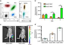
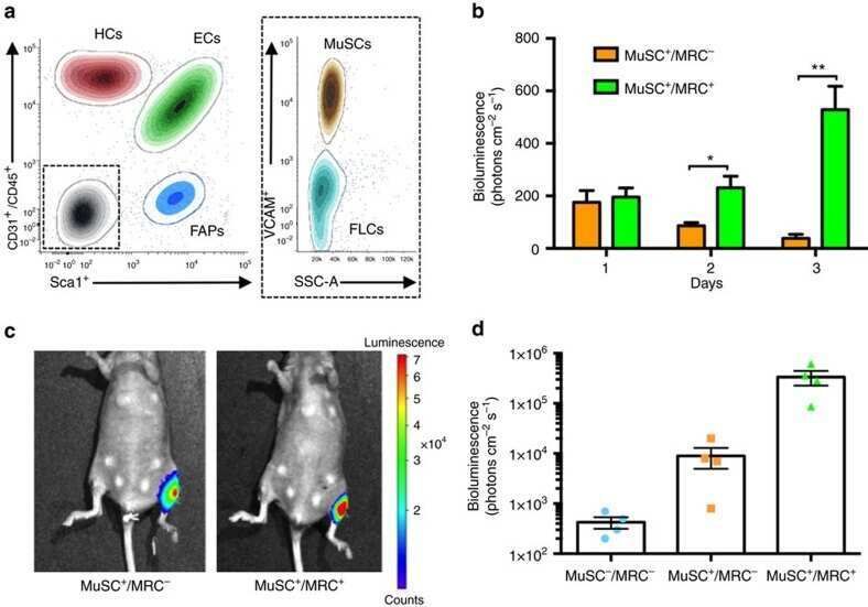
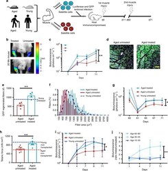
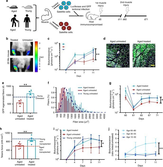
- Fig. 4 Transient OSKMNL expression restores aged muscle stem cell potency. a Schematic showing the experimental design of partially reprogrammed aged mouse and human MuSCs. b Representative images of bioluminescence measured from mice 11 days after transplantation and injury in TiA muscles of treated/untreated Luciferase + mouse MuSCs. c Quantified results of bioluminescence in b at different time points following transplantation and injury ( n = 10). d Representative immunofluorescence of GFP expression in TiA muscle cross-sections of mice imaged and quantified in c and d , isolated 11 days after transplantation (Scale bar = 500 mum). e Quantification of immunofluorescence staining in d ( n = 5). f Quantification of the cross-sectional area of donor-derived GFP + fibers in TiA muscles that were recipients of transplanted MuSCs ( n = 5). g Results of bioluminescence imaging of TiA muscles reinjured after 60 days (second injury) after MuSC transplantations ( n = 6). The second injury was performed to test whether the bioluminescence signal increased as a consequence of activating and expanding luciferase + /GFP + MuSCs that were initially transplanted and that had engrafted under the basal lamina. h Tetanic force measurements of aged muscles injured and transplanted with aged MuSCs. TiA muscles were dissected and electrophysiology ex vivo for tetanic measurement performed. Baseline of force production of untransplanted muscles was measured in young (4 months, blue broken line)
- Conjugate
- Green dye
- Submitted by
- Invitrogen Antibodies (provider)
- Main image
- Experimental details
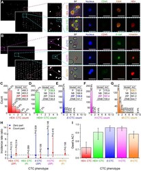
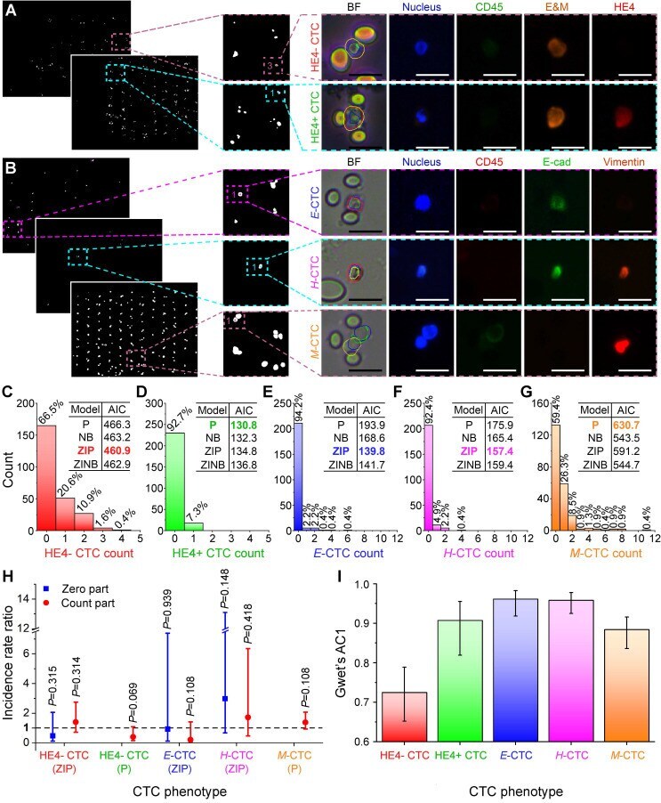
- Figure 4 Benchmarking ALICE CTC phenotypic count against human enumeration in real fluorescent images. Identification, localization and enumeration of CTC phenotypes: (A) HE4- (DAPI+/CD45-/E&M+/HE4-) and HE4+ (DAPI+/CD45-/E&M+/HE4+) CTCs from 61 ovarian cancer patients, (B) E CTCs (DAPI+/CD45-/E-cadherin+/vimentin-), H CTCs (DAPI+/CD45-/E-cadherin+/vimentin+) and M CTCs (DAPI+/CD45-/E-cadherin-/vimentin+) from 46 pancreatic cancer patients. E&M denotes combined epithelial and mesenchymal markers. Scale bar: 20 um. (C-G) Distribution of the phenotypic count for HE4- CTC, HE4+ CTC, E -CTC, H -CTC and M -CTC. Inset tables show the AIC values for the 4 fitted regression models: Poisson (P), negative binomial (NB), zero-inflated Poisson (ZIP) and zero-inflated negative binomial (ZINB) model and the model with the lowest AIC value is bolded and colored. (H) Incidence rate ratio (IRR) plot indicating the CTC phenotypic counts of ALICE and human are statistically indifferent. The fitted regression models are listed for each CTC phenotypes and the zero-inflated models have a zero part and a count part whereas nonzero-inflated models only have a count part. The dash line represents IRR=1 and error bars denote the 95% CI of the IRR. (I) Agreement analysis between ALICE and human counts using Gwet's AC1 for the 5 CTC phenotypes. Error bars represent the 95% CI.
- Conjugate
- Green dye
- Submitted by
- Invitrogen Antibodies (provider)
- Main image

- Experimental details
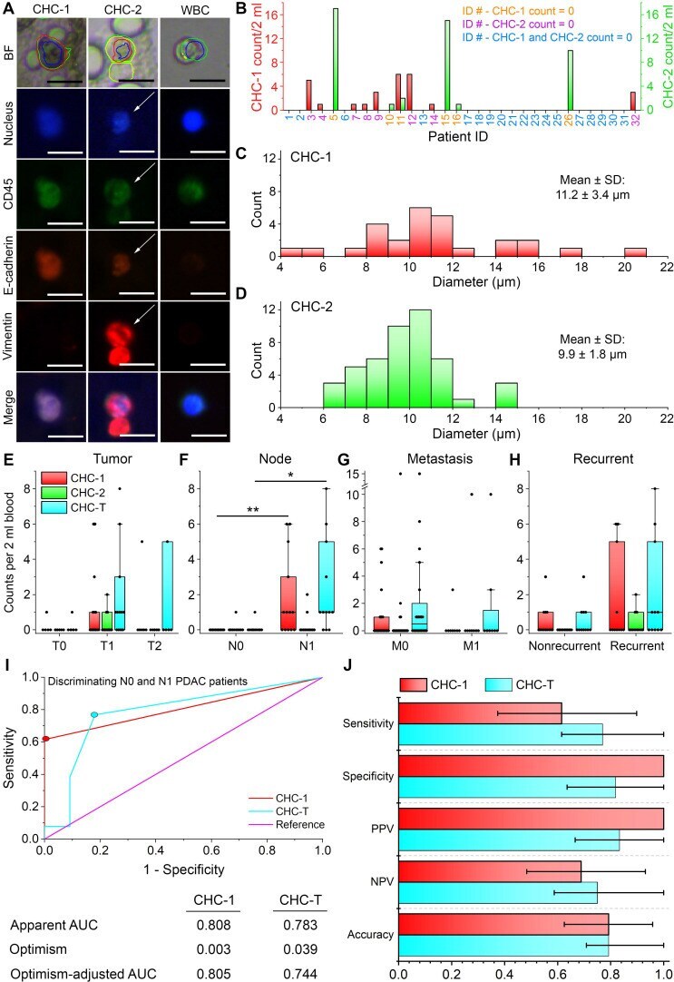
- Figure 5 Circulating hybrid cells (CHCs) in pancreatic cancer patients. (A) Two populations of fusion hybrid identified by ALICE: CHC-1 (DAPI+/CD45+/E-cadherin+/vimentin-) and CHC-2 (DAPI+/CD45+/E-cadherin+/vimentin+) embedded in an overwhelming population of WBCs (DAPI+/CD45+/E-cadherin-/vimentin-) in pancreatic cancer patients. Scale bar: 20 um. (B) Frequency histogram of CHC-1 and CHC-2 counts in pancreatic cancer patients. (C-D) Size distribution of CHC-1 and CHC-2. (E-H) Correlation of CHC-1, CHC-2 and CTC-T with T stage ( n =24), N stage ( n =24), M stage ( n =32) and recurrence ( n =32). * - P < 0.05; ** - P < 0.01 from Mann-Whitney U test. (I) Receiver operating characteristic (ROC) curves for CHC-1 and CHC-T in differentiating N0 and N1 PDAC patients with their respective apparent area under the curve (AUC), optimism and optimism-adjusted AUC calculated over 10000 bootstrap iterations. Colored dots represent the selected cutoff of 1 CHC-1/2 ml of blood and 1 CHC-T/2 ml of blood. (J) Validity of CHC-1 and CHC-T as PDAC node-positive biomarker in terms of the sensitivity, specificity, positive predicted value (PPV), negative predicted value (NPV) and accuracy. The error bars denote the 95% CI.
- Conjugate
- Green dye
- Submitted by
- Invitrogen Antibodies (provider)
- Main image
- Experimental details
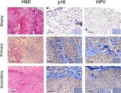
- Fig 3 Immunohistological features of serially transplanted cervical cancer PDXs. Representative example of a PDX showing comparable histology (H&E) and immunoreactivity for diagnostic markers of cervical cancer in the primary biopsy and primary and secondary PDXs. Columns 2 and 3 show staining patterns for p16 INK4a (brown nuclear staining), HPV (brown nuclear and cytoplasmic immunostaining). Insets, isotype IgG control showing negative immunostaining. Scale bars 10 mum.
- Conjugate
- Green dye
- Submitted by
- Invitrogen Antibodies (provider)
- Main image
- Experimental details
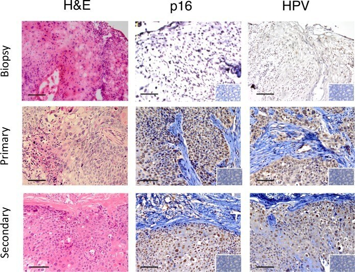
- Fig. 3 Cell-line validation study. (a) Immunofluorescence staining of 5 pancreatic cancer cell lines: AsPC-1, CFPAC-1, MIAPaCa-2, BxPC-3 and Panc-1 with EpCAM and pan-CK. (b) Quantitative comparison of the fluorescence intensity of EpCAM for the 5 pancreatic cancer cell lines. (c) Immunofluorescence staining of the 5 cell lines with E -cad, vimentin, CD45 and DAPI showing the varying epithelial/mesenchymal expressions of the cell lines. Scale bar: 20 mum. Note: A.U. denotes arbitrary unit. The error bars represent the standard deviation. Fig. 3
- Conjugate
- Green dye

- Submitted by
- Invitrogen Antibodies (provider)
- Main image
- Experimental details
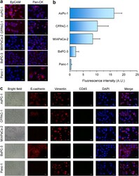
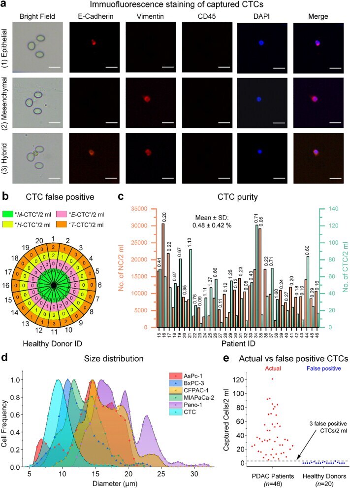
- Fig. 4 CTC characterization: phenotype identification, false positive, capture purity, cell-size distribution and actual CTC count versus false positive CTC count. (a) Immunofluorescence staining of captured CTCs: (1) epithelial CTCs (DAPI+/CD45-/E-cad+/vimentin-), (2) mesenchymal CTCs (DAPI+/CD45-/E-cad-/vimentin+), (3) hybrid CTCs (DAPI+/CD45-/E-cad+/vimentin+). Scale bar: 20 mum. (b) False positive CTC counts from healthy donors ( n = 20). The outer most ring of numbers refers to the healthy donor ID and the inner band of rings indicate the no. of false positive CTC phenotypes. A maximum of 3 false positive CTCs/2 ml was obtained. (c) CTC capture purity as measured by the number of captured WBCs from PDAC patients ( n = 32). The figure above each individual bar denotes the CTC/WBC percentage ratio for a patient. (d) CTC size distribution: captured CTCs versus the 5 pancreatic cancer cell lines (AsPc-1, BxPC-3, CFPAC-1, MIAPaCa-2 and Panc-1). (e) Comparing actual CTC count from PDAC patients ( n = 46) with false positive CTC count from healthy donors ( n = 20) for a proper handling of the false positive issue. The dashed line represents the detection noise level at 3 false positive CTCs/2 ml of blood. Fig. 4
- Conjugate
- Green dye
- Submitted by
- Invitrogen Antibodies (provider)
- Main image
- Experimental details
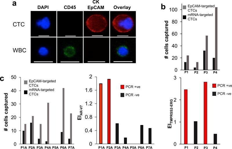
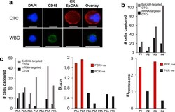
- Figure 4 Analysis of clinical samples ( A ) Representative image of a CTC captured from a prostate cancer patient blood sample versus a white blood cell (WBC). The cells were stained with APC-labeled anti-CK, APC-labeled anti-EpCAM, AF488-labeled anti-CD45, and DAPI. Only CK + /EpCAM + /CD45 - /DAPI + cells are counted as CTC. The scale bar is 15 um. ( B ) Analysis of blood samples collected from prostate cancer patients for the TMPRSS2-ERG gene fusion. Samples that tested positive for the gene fusion (see Supplementary Figure 11 ) exhibited significantly higher expression indices than those that tested negative. ( C ) Analysis of blood samples collected from prostate cancer patients for the androgen receptor splice variant AR-V7. Samples that tested positive for AR-V7 (see Supplementary Figure 12 ) exhibited significantly higher expression indices than those that tested negative.
- Conjugate
- Green dye