Explore
 Validate
 Learn
Learn Flow cytometry
Antibody data
- Antibody Data
- Antigen structure
- References [15]
- Comments [0]
- Validations
- Flow cytometry [2]
- Other assay [3]
Submit
Validation data
Reference
Comment
Report error
- Product number
- 12-0457-42 - Provider product page

- Provider
- Invitrogen Antibodies
- Product name
- CD45RO Monoclonal Antibody (UCHL1), PE, eBioscience™
- Antibody type
- Monoclonal
- Antigen
- Other
- Description
- Description: The UCHL1 monoclonal antibody reacts with human CD45RO, a 180 kDa isoform of CD45. CD45RO is expressed by most thymocytes, activated memory T cells, granulocytes and monocytes. CD22 is a ligand for CD45RO. Expression of CD45RO and CD45RA is used commonly to discriminate subsets of peripheral T cells. Applications Reported: The UCHL1 antibody has been reported for use in flow cytometric analysis. Applications Tested: This UCHL1 antibody has been pre-titrated and tested by flow cytometric analysis of normal human peripheral blood cells. This can be used at 5 µL (0.5 µg) per test. A test is defined as the amount (µg) of antibody that will stain a cell sample in a final volume of 100 µL. Cell number should be determined empirically but can range from 10^5 to 10^8 cells/test. Excitation: 488-561 nm; Emission: 578 nm; Laser: Blue Laser, Green Laser, Yellow-Green Laser. Filtration: 0.2 µm post-manufacturing filtered.
- Reactivity
- Human
- Host
- Mouse
- Conjugate
- Yellow dye
- Isotype
- IgG
- Antibody clone number
- UCHL1
- Vial size
- 100 Tests
- Concentration
- 5 µL/Test
- Storage
- 4° C, store in dark, DO NOT FREEZE!
Submitted references Development of CAR-T cell therapy for B-ALL using a point-of-care approach.
Longitudinal analysis of subtype C envelope tropism for memory CD4(+) T cell subsets over the first 3 years of untreated HIV-1 infection.
Clinically compliant spatial and temporal imaging of chimeric antigen receptor T-cells.
Modulation of Endoplasmic Reticulum Stress Controls CD4(+) T-cell Activation and Antitumor Function.
Blocking the recruitment of naive CD4(+) T cells reverses immunosuppression in breast cancer.
Effector and Central Memory Poly-Functional CD4(+) and CD8(+) T Cells are Boosted upon ZOSTAVAX(®) Vaccination.
Somatic Variation of T-Cell Receptor Genes Strongly Associate with HLA Class Restriction.
Functional expression of CD137 (4-1BB) on T helper follicular cells.
Delta-like 1-mediated Notch signaling enhances the in vitro conversion of human memory CD4 T cells into FOXP3-expressing regulatory T cells.
Expression of the memory marker CD45RO on helper T cells in macaques.
Dual role of miR-21 in CD4+ T-cells: activation-induced miR-21 supports survival of memory T-cells and regulates CCR7 expression in naive T-cells.
Signal transducer and activator of transcription 3 (STAT3) mutations underlying autosomal dominant hyper-IgE syndrome impair human CD8(+) T-cell memory formation and function.
CpG and non-CpG oligodeoxynucleotides directly costimulate mouse and human CD4+ T cells through a TLR9- and MyD88-independent mechanism.
Longevity and composition of cellular immune responses following experimental Plasmodium falciparum malaria infection in humans.
Impaired CD4 and CD8 effector function and decreased memory T cell populations in ICOS-deficient patients.
de Macedo Abdo L, Barros LRC, Saldanha Viegas M, Vieira Codeço Marques L, de Sousa Ferreira P, Chicaybam L, Bonamino MH
Oncoimmunology 2020;9(1):1752592
Oncoimmunology 2020;9(1):1752592
Longitudinal analysis of subtype C envelope tropism for memory CD4(+) T cell subsets over the first 3 years of untreated HIV-1 infection.
Gartner MJ, Gorry PR, Tumpach C, Zhou J, Dantanarayana A, Chang JJ, Angelovich TA, Ellenberg P, Laumaea AE, Nonyane M, Moore PL, Lewin SR, Churchill MJ, Flynn JK, Roche M
Retrovirology 2020 Aug 6;17(1):24
Retrovirology 2020 Aug 6;17(1):24
Clinically compliant spatial and temporal imaging of chimeric antigen receptor T-cells.
Emami-Shahri N, Foster J, Kashani R, Gazinska P, Cook C, Sosabowski J, Maher J, Papa S
Nature communications 2018 Mar 14;9(1):1081
Nature communications 2018 Mar 14;9(1):1081
Modulation of Endoplasmic Reticulum Stress Controls CD4(+) T-cell Activation and Antitumor Function.
Thaxton JE, Wallace C, Riesenberg B, Zhang Y, Paulos CM, Beeson CC, Liu B, Li Z
Cancer immunology research 2017 Aug;5(8):666-675
Cancer immunology research 2017 Aug;5(8):666-675
Blocking the recruitment of naive CD4(+) T cells reverses immunosuppression in breast cancer.
Su S, Liao J, Liu J, Huang D, He C, Chen F, Yang L, Wu W, Chen J, Lin L, Zeng Y, Ouyang N, Cui X, Yao H, Su F, Huang JD, Lieberman J, Liu Q, Song E
Cell research 2017 Apr;27(4):461-482
Cell research 2017 Apr;27(4):461-482
Effector and Central Memory Poly-Functional CD4(+) and CD8(+) T Cells are Boosted upon ZOSTAVAX(®) Vaccination.
Sei JJ, Cox KS, Dubey SA, Antonello JM, Krah DL, Casimiro DR, Vora KA
Frontiers in immunology 2015;6:553
Frontiers in immunology 2015;6:553
Somatic Variation of T-Cell Receptor Genes Strongly Associate with HLA Class Restriction.
Klarenbeek PL, Doorenspleet ME, Esveldt RE, van Schaik BD, Lardy N, van Kampen AH, Tak PP, Plenge RM, Baas F, de Bakker PI, de Vries N
PloS one 2015;10(10):e0140815
PloS one 2015;10(10):e0140815
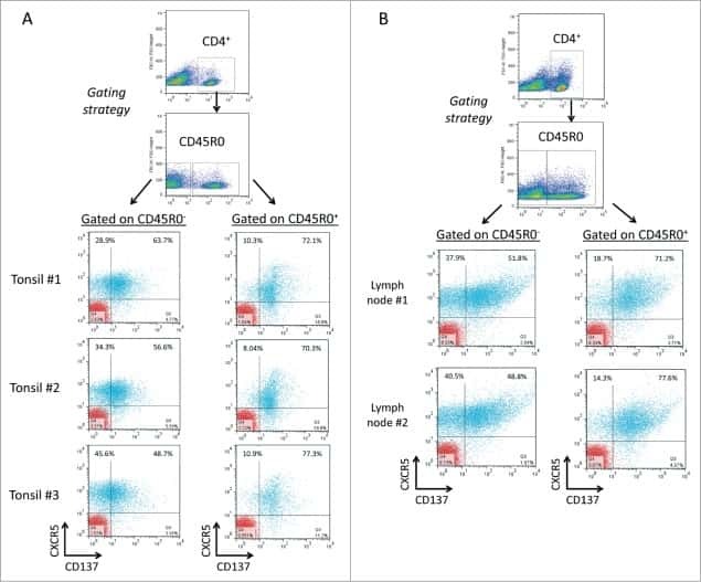
Functional expression of CD137 (4-1BB) on T helper follicular cells.
Alfaro C, Echeveste JI, Rodriguez-Ruiz ME, Solorzano JL, Perez-Gracia JL, Idoate MA, Lopez-Picazo JM, Sanchez-Paulete AR, Labiano S, Rouzaut A, Oñate C, Aznar A, Lozano MD, Melero I
Oncoimmunology 2015 Dec;4(12):e1054597
Oncoimmunology 2015 Dec;4(12):e1054597
Delta-like 1-mediated Notch signaling enhances the in vitro conversion of human memory CD4 T cells into FOXP3-expressing regulatory T cells.
Mota C, Nunes-Silva V, Pires AR, Matoso P, Victorino RM, Sousa AE, Caramalho I
Journal of immunology (Baltimore, Md. : 1950) 2014 Dec 15;193(12):5854-62
Journal of immunology (Baltimore, Md. : 1950) 2014 Dec 15;193(12):5854-62
Expression of the memory marker CD45RO on helper T cells in macaques.
Valentine M, Song K, Maresh GA, Mack H, Huaman MC, Polacino P, Ho O, Cristillo A, Kyung Chung H, Hu SL, Pincus SH
PloS one 2013;8(9):e73969
PloS one 2013;8(9):e73969
Dual role of miR-21 in CD4+ T-cells: activation-induced miR-21 supports survival of memory T-cells and regulates CCR7 expression in naive T-cells.
Smigielska-Czepiel K, van den Berg A, Jellema P, Slezak-Prochazka I, Maat H, van den Bos H, van der Lei RJ, Kluiver J, Brouwer E, Boots AM, Kroesen BJ
PloS one 2013;8(10):e76217
PloS one 2013;8(10):e76217
Signal transducer and activator of transcription 3 (STAT3) mutations underlying autosomal dominant hyper-IgE syndrome impair human CD8(+) T-cell memory formation and function.
Ives ML, Ma CS, Palendira U, Chan A, Bustamante J, Boisson-Dupuis S, Arkwright PD, Engelhard D, Averbuch D, Magdorf K, Roesler J, Peake J, Wong M, Adelstein S, Choo S, Smart JM, French MA, Fulcher DA, Cook MC, Picard C, Durandy A, Tsumura M, Kobayashi M, Uzel G, Casanova JL, Tangye SG, Deenick EK
The Journal of allergy and clinical immunology 2013 Aug;132(2):400-11.e9
The Journal of allergy and clinical immunology 2013 Aug;132(2):400-11.e9
CpG and non-CpG oligodeoxynucleotides directly costimulate mouse and human CD4+ T cells through a TLR9- and MyD88-independent mechanism.
Landrigan A, Wong MT, Utz PJ
Journal of immunology (Baltimore, Md. : 1950) 2011 Sep 15;187(6):3033-43
Journal of immunology (Baltimore, Md. : 1950) 2011 Sep 15;187(6):3033-43
Longevity and composition of cellular immune responses following experimental Plasmodium falciparum malaria infection in humans.
Teirlinck AC, McCall MB, Roestenberg M, Scholzen A, Woestenenk R, de Mast Q, van der Ven AJ, Hermsen CC, Luty AJ, Sauerwein RW
PLoS pathogens 2011 Dec;7(12):e1002389
PLoS pathogens 2011 Dec;7(12):e1002389
Impaired CD4 and CD8 effector function and decreased memory T cell populations in ICOS-deficient patients.
Takahashi N, Matsumoto K, Saito H, Nanki T, Miyasaka N, Kobata T, Azuma M, Lee SK, Mizutani S, Morio T
Journal of immunology (Baltimore, Md. : 1950) 2009 May 1;182(9):5515-27
Journal of immunology (Baltimore, Md. : 1950) 2009 May 1;182(9):5515-27
No comments: Submit comment
Supportive validation
- Submitted by
- Invitrogen Antibodies (provider)
- Main image
- Experimental details
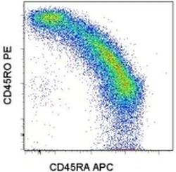
- Staining of normal human peripheral blood cells with Anti-Human CD45RA APC (Product # 17-0458-42) and Anti-Human CD45RO PE. Cells in the lymphocyte gate were used for analysis.
- Conjugate
- Yellow dye

- Submitted by
- Invitrogen Antibodies (provider)
- Main image

- Experimental details
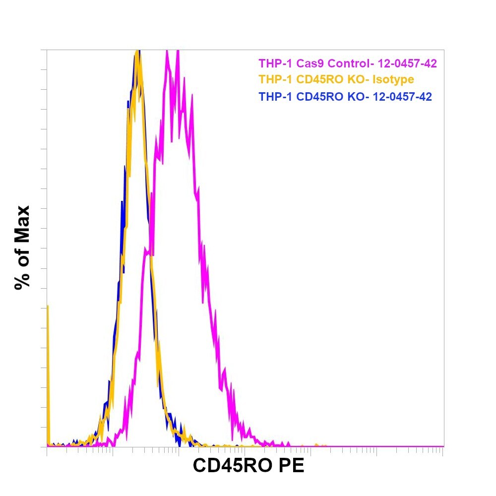
- Knockout of CD45RO was achieved by CRISPR-Cas9 genome editing using LentiArray™ Lentiviral sgRNA (Product # A32042, Assay ID CRISPR664203_LV) and LentiArray Cas9 Lentivirus (Product # A32064). Flow cytometry analysis of CD45RO was performed by staining THP-1 CD45RO Knock out cells with 0.5 µg Mouse IgG2a kappa Isotype Control (eBM2a), PE, eBioscience™ (Product # 12-4724-82, yellow histogram) or 0.5 µg CD45RO Monoclonal Antibody (UCHL1), PE, eBioscience™ (Product # 12-0457-42, blue histogram). THP-1 Cas9 control cells were also stained with0.5 µg CD45RO Monoclonal Antibody (UCHL1), PE, eBioscience™ (Product # 12-0457-42, pink histogram). Lossof signal was observed in the KOcells stained with CD45RO antibody clone UCHL1 but not in the control Cas9cells. Viable cells were used for analysis, as determined by Fixable Viability DyeeFluor™780 (Product # 65-0865-18).
- Conjugate
- Yellow dye
Supportive validation
- Submitted by
- Invitrogen Antibodies (provider)
- Main image
- Experimental details
- NULL
- Conjugate
- Yellow dye
- Submitted by
- Invitrogen Antibodies (provider)
- Main image
- Experimental details
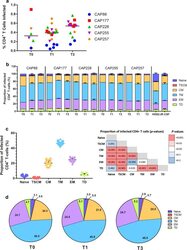
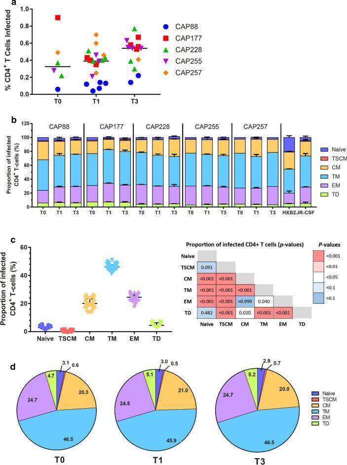
- Fig. 3 Transitional memory and effector memory cells were most frequently infected by C-HIV Envs. a Each data point represents the percentage of infected CD4 + T cells with one pseudovirus (averaged from four independent seronegative blood bank donors). The Env donor is indicated as follows; CAP88 (blue circles), CAP177 (red squares), CAP228 (green triangles), CAP255 (purple inverted triangles) and CAP257 (orange diamonds). Black lines represents the median of all pseudoviruses within each time point. Comparisons were made using a Kruskal-Wallis test with Dunn's post hoc test for multiple comparisons. b Stacked bar graphs represent the contribution of each T cell subset to the pool of infected CD4 + T cells. Values represent the median percentage of infected CD4 + T-cells (averaged across four HIV-seronegative PBMC donors) that belong to the indicated subset [naive; dark blue, T stem cell memory (TSCM); red, central memory (CM); yellow, transitional memory (TM); light blue, effector memory (EM); purple and terminally differentiated (TD); green], and are stratified by participant and time point. Error bars represent the interquartile range. c Dot plot representing the proportion of each T cell subset contributing to the total pool of infected cells for all Env-pseudoviruses. Each point represents a single virus averaged across four seronegative donors, lines represent median and error bars represent interquartile range. Comparisons were made using a Kruskal-Wallis test with Du
- Conjugate
- Yellow dye
- Submitted by
- Invitrogen Antibodies (provider)
- Main image
- Experimental details
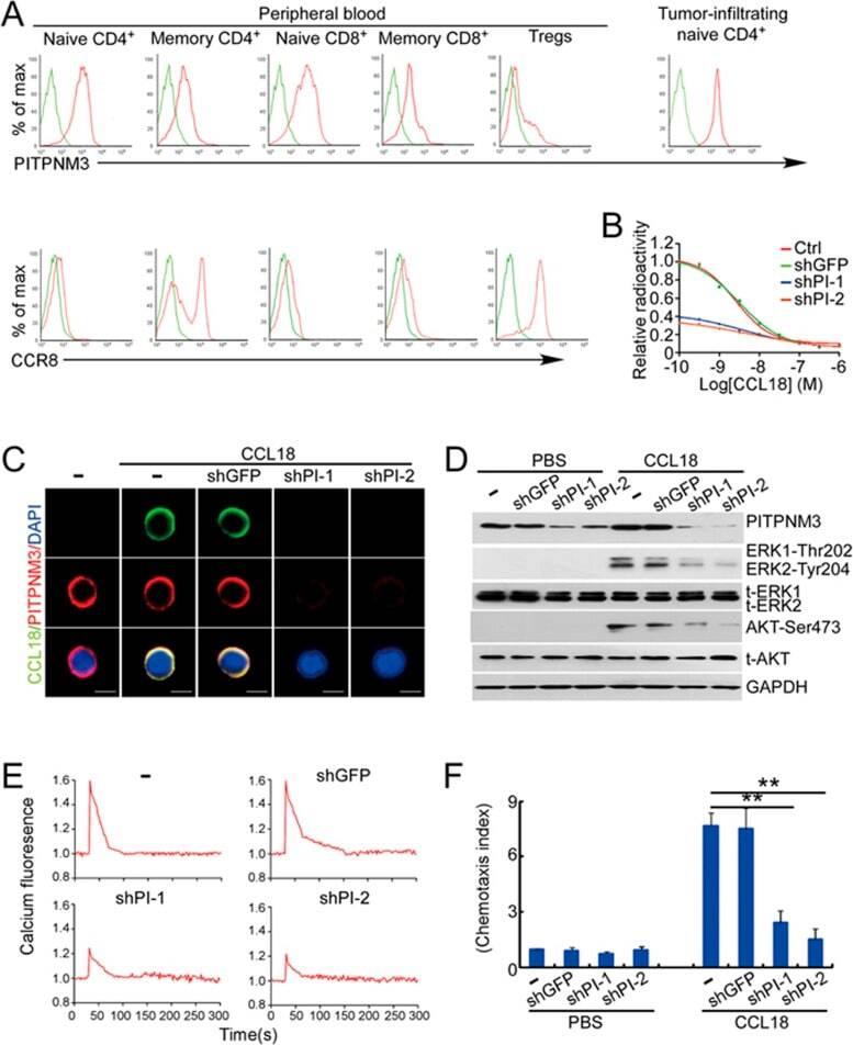
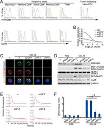
- Figure 5 PITPNM3 is a CCL18 receptor on naive CD4 + T cells. (A) Representative flow cytometry staining for PITPNM3 and CCR8, potential CCL18 receptors, on gated PB T cell subsets and paired TI naive CD4 + T cells of a breast cancer patient. Cells were gated on CD3 + CD45RA + CD45RO - CD25 - CD4 + /CD8 + for naive CD4 + /CD8 + T cells, CD3 + CD45RA - CD45RO + CD25 - CD4 + /CD8 + for memory CD4 + /CD8 + T cells and CD3 + CD4 + CD25 + for Tregs). Quantitation of PITPNM3 and CCR8 expression on T cell subsets for eight breast cancer patients is provided in Supplementary information, Figure S8A . (B-F) Knockdown of PITPNM3 in naive CD4 + T cells inhibits CCL18 binding, signaling and chemotaxis. (B) Binding of 125 I-CCL18 to naive CD4 + T cells, knocked down or not for PITPNM3 (shPI-1,2) in the presence of increasing concentrations of unlabeled CCL18. Shown are the representative assays for three independent experiments using PB T cells from three normal donors. (C) Representative fluorescence microscopy images of CCL18 binding to naive CD4 + T cells, knocked down or not for PITPNM3 , stained for PITPNM3 and CCL18 3 h after adding CCL18. Scale bar, 5 mum. Shown are the representative images for three independent experiments using PB T cells from three normal donors. (D) Immunoblot of CCL18-treated naive CD4 + T cells, knocked down or not for PITPNM3 , showing expression of PITPNM3 and phosphorylated/total (t-) Erk1/2 and Akt, relative to GAPDH as a loading control. Blots are repres
- Conjugate
- Yellow dye