 Explore
 Validate
 Learn
Learn Flow cytometry
Antibody data
- Antibody Data
- Antigen structure
- References [3]
- Comments [0]
- Validations
- Flow cytometry [2]
- Other assay [1]
Submit
Validation data
Reference
Comment
Report error
- Product number
- 12-9979-42 - Provider product page

- Provider
- Invitrogen Antibodies
- Product name
- CD45RA Monoclonal Antibody (JS-83), PE, eBioscience™
- Antibody type
- Monoclonal
- Antigen
- Other
- Description
- Description: The JS-83 monoclonal antibody reacts with human CD45RA, a 220 kDa molecule expressed by subpopulations of CD4^+ peripheral T lymphocytes, CD8^+ peripheral T lymphocytes, and B cells. The CD45RA^+ T cell populations are mainly naive/virgin allowing the use of JS-83 mAb as a phenotypic marker to discriminate T cell subsets. Applications Reported: JS-83 has been reported for use in flow cytometric analysis. Applications Tested: This JS-83 antibody has been pre-titrated and tested by flow cytometric analysis of normal human peripheral blood cells. This can be used at 5 µL (0.03 µg) per test. A test is defined as the amount (µg) of antibody that will stain a cell sample in a final volume of 100 µL. Cell number should be determined empirically but can range from 10^5 to 10^8 cells/test. Excitation: 488-561 nm; Emission: 578 nm; Laser: Blue Laser, Green Laser, Yellow-Green Laser. Filtration: 0.2 µm post-manufacturing filtered.
- Reactivity
- Human
- Host
- Mouse
- Conjugate
- Yellow dye
- Isotype
- IgG
- Antibody clone number
- JS-83
- Vial size
- 100 Tests
- Concentration
- 5 µL/Test
- Storage
- 4° C, store in dark, DO NOT FREEZE!
Submitted references IL-32γ potentiates tumor immunity in melanoma.
Human immunodeficiency virus type I-specific CD8+ T cell subset abnormalities in chronic infection persist through effective antiretroviral therapy.
TCR-induced downregulation of protein tyrosine phosphatase PEST augments secondary T cell responses.
Gruber T, Kremenovic M, Sadozai H, Rombini N, Baeriswyl L, Maibach F, Modlin RL, Gilliet M, von Werdt D, Hunger RE, Seyed Jafari SM, Parisi G, Abril-Rodriguez G, Ribas A, Schenk M
JCI insight 2020 Sep 17;5(18)
JCI insight 2020 Sep 17;5(18)
Human immunodeficiency virus type I-specific CD8+ T cell subset abnormalities in chronic infection persist through effective antiretroviral therapy.
Pohling J, Zipperlen K, Hollett NA, Gallant ME, Grant MD
BMC infectious diseases 2010 May 25;10:129
BMC infectious diseases 2010 May 25;10:129
TCR-induced downregulation of protein tyrosine phosphatase PEST augments secondary T cell responses.
Arimura Y, Vang T, Tautz L, Williams S, Mustelin T
Molecular immunology 2008 Jun;45(11):3074-84
Molecular immunology 2008 Jun;45(11):3074-84
No comments: Submit comment
Supportive validation
- Submitted by
- Invitrogen Antibodies (provider)
- Main image

- Experimental details
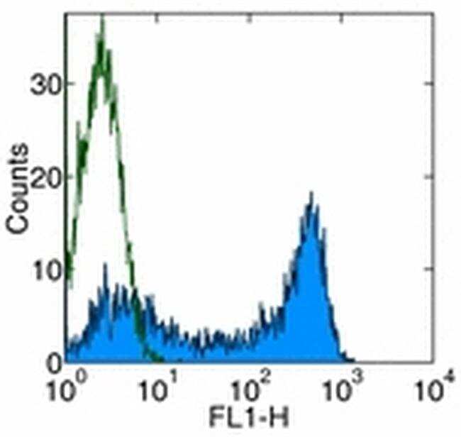
- Surface staining of normal human peripheral blood cells with Anti-Human CD45RA FITC. Appropriate isotype controls were used (open histogram). Cells in the lymphocyte population were used for analysis.
- Conjugate
- Yellow dye
- Submitted by
- Invitrogen Antibodies (provider)
- Main image

- Experimental details
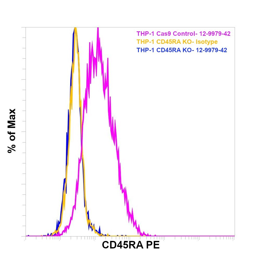
- Knockout of CD45RA was achieved by CRISPR-Cas9 genome editing using LentiArray™ Lentiviral sgRNA (Product # A32042, Assay ID CRISPR664203_LV) and LentiArray Cas9 Lentivirus (Product # A32064). Flow cytometry analysis of CD45RA was performed by staining THP-1 CD45RA Knock out cells with 0.03 µg Mouse IgG1 kappa Isotype Control (P3.6.2.8.1), PE, eBioscience™ (Product # 12-4714-82) or 0.03 µg CD45RA Monoclonal Antibody (JS-83), PE, eBioscience™ (Product # 12-9979-42, blue histogram). THP-1 Cas9 control cells were also stained with0.03 µg CD45RA Monoclonal Antibody (JS-83), PE, eBioscience™ (Product # 12-9979-42, pink histogram). Lossof signal was observed in the KOcells stained with CD45RA antibody clone JS-83 but not in the control Cas9cells. Viable cells were used for analysis, as determined by Fixable Viability DyeeFluor™780 (Product # 65-0865-18).
- Conjugate
- Yellow dye
Supportive validation
- Submitted by
- Invitrogen Antibodies (provider)
- Main image
- Experimental details
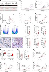
- Figure 4 IL-32 induces a systemic CD8 + T cell-mediated tumor-specific immune response. ( A ) Experimental setup for in vivo tumor treatments. MC38 and B16F10 were inoculated in C57BL/6J mice, and 4T1 tumors in BALB/c mice ( A - O ). ( B ) Growth curves of IL-32-, cGAMP-, or PBS-treated primary and ( C ) contralateral, nontreated B16F10 melanomas. * P < 0.05, ** P < 0.01, *** P < 0.001. Data are representative of 4 independent experiments, with n = 6 mice per group. ( D ) Kaplan-Meier survival curves of B16F10-bearing mice treated with IL-32 ( n = 6) or PBS ( n = 8). ( E ) Representative growth curves of IL-32-treated and untreated MC38 colon adenocarcinoma ( n = 6) and ( F ) orthotopic 4T1 breast tumors ( n = 10). ( G-O ) On day 14, the primary treated tumors and spleens were harvested for flow cytometric analyses, IHC or TCRbeta chain sequencing. ( G ) Representative flow cytometry plots displaying frequencies of CD45 + immune cells for each treatment group and ( H ) their quantification shown as relative frequencies ( n = 18). ( I ) Relative frequencies of CD8 + and CD4 + T cells as a percentage of live cells ( n = 18). ( J ) Representative immunohistochemical staining and ( K ) morphometric enumeration as cells/mm 2 of CD8 + T cells ( n = 3). Scale bar: 20 mum. ( L ) Relative frequencies of IFN-gamma + cells as percentage of CD8 + T cells ( n = 12). ( M ) Frequencies of Nur77-GFP + cells of CD8 + T cells, as determined by flow cytometry in B16F10-inoculated and PBS- ( n =
- Conjugate
- Yellow dye
