 Explore
 Validate
 Learn
Learn Western blot
 Immunohistochemistry
Antibody data
- Antibody Data
- Antigen structure
- References [5]
- Comments [0]
- Validations
- Immunohistochemistry [1]
- Flow cytometry [1]
Submit
Validation data
Reference
Comment
Report error
- Product number
- 14-9458-82 - Provider product page

- Provider
- Invitrogen Antibodies
- Product name
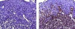
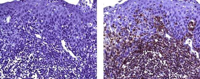
- CD45RB Monoclonal Antibody (PD7/26), eBioscience™
- Antibody type
- Monoclonal
- Antigen
- Other
- Description
- Description: The monoclonal antibody PD7/26 recognizes CD45RB, which is an isoform of the Leukocyte Common Antigen (LCA) also known as CD45. CD45RB is a protein tyrosine phosphatase receptor that is present on most hematopoietic cells and absent on non-hematopoietic cells. Expression of CD45RB is more restricted than CD45 and can only be found on T cell subsets, B, NK, and myeloid cells. Regulatory T cells (Tregs) have been show to express low levels of CD45RB, which may correlate with increased migration to sites of infection. In contrast, high CD45RB expression results in IL-2 and IFN gamma production instead of IL-4 in vitro. Applications Reported: This PD7/26 antibody has been reported for use in Flow Cytometric Analysis, Western Blotting, Immunohistochemical Staining of Formalin-Fixed Paraffin Embedded Tissue Sections, and Immunocytochemistry. Applications Tested: This PD7/26 antibody has been tested by immunohistochemistry on formalin-fixed paraffin embedded human tonsil using low pH antigen retrieval. This can be used at less than or equal to 5 µg/mL. This PD7/26 antibody has also been tested by western blot at 5 µg/mL and and by flow cytometric analysis on human peripheral blood cells at 0.25 µg/test. A test is defined as the amount (µg) of antibody that will stain a cell sample in a final volume of 100 µL. Cell number should be determined empirically but can range from 10^5 to 10^8 cells/test. It is recommended that the antibody be carefully titrated for optimal performance in the assay of interest. Purity: Greater than 90%, as determined by SDS-PAGE. Aggregation: Less than 10%, as determined by HPLC. Filtration: 0.2 µm post-manufacturing filtered.
- Reactivity
- Human
- Host
- Mouse
- Isotype
- IgG
- Antibody clone number
- PD7/26
- Vial size
- 100 µg
- Concentration
- 0.5 mg/mL
- Storage
- 4° C
Submitted references Inflammatory malignant fibrous histiocytoma: distinction from Hodgkin's disease and non-Hodgkin's lymphoma by a panel of leukocyte markers.
CD45 isoform expression on human haemopoietic cells at different stages of development.
CD45RB expression defines two interconvertible subsets of human CD4+ T cells with memory function.
Isoform-specific associations of CD45 with accessory molecules in human T lymphocytes.
Comparative biochemical and tissue distribution study of four distinct CD45 antigen specificities.
Khalidi HS, Singleton TP, Weiss SW
Modern pathology : an official journal of the United States and Canadian Academy of Pathology, Inc 1997 May;10(5):438-42
Modern pathology : an official journal of the United States and Canadian Academy of Pathology, Inc 1997 May;10(5):438-42
CD45 isoform expression on human haemopoietic cells at different stages of development.
Craig W, Poppema S, Little MT, Dragowska W, Lansdorp PM
British journal of haematology 1994 Sep;88(1):24-30
British journal of haematology 1994 Sep;88(1):24-30
CD45RB expression defines two interconvertible subsets of human CD4+ T cells with memory function.
Horgan KJ, Tanaka Y, Luce GE, van Seventer GA, Nutman TB, Shaw S
European journal of immunology 1994 May;24(5):1240-3
European journal of immunology 1994 May;24(5):1240-3
Isoform-specific associations of CD45 with accessory molecules in human T lymphocytes.
Dianzani U, Redoglia V, Malavasi F, Bragardo M, Pileri A, Janeway CA Jr, Bottomly K
European journal of immunology 1992 Feb;22(2):365-71
European journal of immunology 1992 Feb;22(2):365-71
Comparative biochemical and tissue distribution study of four distinct CD45 antigen specificities.
Pulido R, Cebrián M, Acevedo A, de Landázuri MO, Sánchez-Madrid F
Journal of immunology (Baltimore, Md. : 1950) 1988 Jun 1;140(11):3851-7
Journal of immunology (Baltimore, Md. : 1950) 1988 Jun 1;140(11):3851-7
No comments: Submit comment
Supportive validation
- Submitted by
- Invitrogen Antibodies (provider)
- Main image
- Experimental details
- Immunohistochemistry on formalin-fixed paraffin embedded human tonsil, using 5 µg/mL of Mouse IgG1 Isotype Control (left) or 5 µg/mL Anti-Human CD45RB Purified (right) followed by Anti-Mouse IgG Biotin, and DAB visualization.Nuclei are counterstained with hematoxylin.
Supportive validation
- Submitted by
- Invitrogen Antibodies (provider)
- Main image

- Experimental details
- Knockout of CD45RB was achieved by CRISPR-Cas9 genome editing using LentiArray™ Lentiviral sgRNA (Product # A32042, Assay ID CRISPR664203_LV) and LentiArray Cas9 Lentivirus (Product # A32064). Flow cytometry analysis of CD45RB was performed by staining THP-1 CD45RB Knock out cells with0.25 µg Mouse IgG1 kappa Isotype Control (P3.6.2.8.1), eBioscience™ (Product # 14-4714-82, yellow histogram) or 0.25 µg CD45RB Monoclonal Antibody (PD7/26), eBioscience™ (Product # 14-9458-82, blue histogram) followed by Goat anti-Mouse IgG (H+L), Superclonal™ Recombinant Secondary Antibody, Alexa Fluor™ Plus 488 (Product # A55058, 1:1000 dilution). THP-1 Cas9 control cells was also stained with0.25 µg CD45RB Monoclonal Antibody (PD7/26), eBioscience™ (Product # 14-9458-82, pink histogram) followed by the secondary antibody. Lossof signal was observed in the CD45RB KOcells stained with CD45RB antibody clone PD7/26 but not in the control Cas9cells. Viable cells were used for analysis, as determined by Fixable Viability Dye eFluor™ 780 (Product # 65-0865-18).
