Explore
 Validate
 Learn
Learn Western blot
Antibody data
- Antibody Data
- Antigen structure
- References [3]
- Comments [0]
- Validations
- Western blot [1]
- Immunohistochemistry [1]
- Flow cytometry [3]
Submit
Validation data
Reference
Comment
Report error
- Product number
- MA5-12496 - Provider product page

- Provider
- Invitrogen Antibodies
- Product name
- CD45RB Monoclonal Antibody (PD7/26)
- Antibody type
- Monoclonal
- Antigen
- Other
- Description
- MA5-12496 targets CD45RB in FACS, Western blot and IHC (P) applications and shows reactivity with Human and Mouse samples. This antibody is not recommended for BAF-3 cells in Western blot applications.
- Antibody clone number
- PD7/26
- Concentration
- 0.2 mg/mL
Submitted references PDGFR-β (+) perivascular cells from infantile hemangioma display the features of mesenchymal stem cells and show stronger adipogenic potential in vitro and in vivo.
Mesenchymal stem cells in infantile hemangioma reside in the perivascular region.
Anti-CD45RO suppresses human immunodeficiency virus type 1 replication in microglia: role of Hck tyrosine kinase and implications for AIDS dementia.
Yuan SM, Guo Y, Zhou XJ, Shen WM, Chen HN
International journal of clinical and experimental pathology 2014;7(6):2861-70
International journal of clinical and experimental pathology 2014;7(6):2861-70
Mesenchymal stem cells in infantile hemangioma reside in the perivascular region.
Yuan SM, Chen RL, Shen WM, Chen HN, Zhou XJ
Pediatric and developmental pathology : the official journal of the Society for Pediatric Pathology and the Paediatric Pathology Society 2012 Jan-Feb;15(1):5-12
Pediatric and developmental pathology : the official journal of the Society for Pediatric Pathology and the Paediatric Pathology Society 2012 Jan-Feb;15(1):5-12
Anti-CD45RO suppresses human immunodeficiency virus type 1 replication in microglia: role of Hck tyrosine kinase and implications for AIDS dementia.
Kim MO, Suh HS, Si Q, Terman BI, Lee SC
Journal of virology 2006 Jan;80(1):62-72
Journal of virology 2006 Jan;80(1):62-72
No comments: Submit comment
Supportive validation
- Submitted by
- Invitrogen Antibodies (provider)
- Main image
- Experimental details
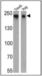
- Western blot analysis of CD45RB was performed by loading 25 µg of Daudi (Lane 1) and Raji (Lane 2) cell lysates onto an SDS polyacrylamide gel. Proteins were transferred to a PVDF membrane and blocked at 4ºC overnight. The membrane was probed with a CD45RB monoclonal antibody (Product # MA5-12496) at a dilution of 1:10000 (Daudi) and 1:500 (Raji) overnight at 4°C, washed in TBST, and probed with an HRP-conjugated secondary antibody for 1 hr at room temperature in the dark. Chemiluminescent detection was performed using Pierce ECL Plus Western Blotting Substrate (Product # 32132). Results show a band at approx. 240 kDa.

Supportive validation
- Submitted by
- Invitrogen Antibodies (provider)
- Main image
- Experimental details
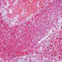
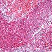
- Formalin-fixed, paraffin-embedded human tonsil stained with CD45RB antibody using peroxidase-conjugate and AEC chromogen. Note cell membrane staining of B lymphocytes.
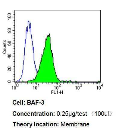
Supportive validation
- Submitted by
- Invitrogen Antibodies (provider)
- Main image
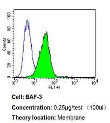
- Experimental details
- Flow cytometry analysis of CD45RB in BAF-3 cells compared to an isotype control (blue). Cells were harvested, adjusted to a concentration of 1-5x10^6 cells/mL, fixed with 2% paraformaldehyde, washed with PBS, and incubated with CD45RB monoclonal antibody (Product # MA5-12496) at a dilution of 0.25 µg/test for 60 min at room temperature. Cells were then blocked in a solution of 2% BSA-PBS for 30 min at room temperature, incubated for 40 min at room temperature in the dark using a Dylight 488-conjugated goat anti-mouse IgG (H+L) secondary antibody, and re-suspended in PBS for FACS analysis.
- Submitted by
- Invitrogen Antibodies (provider)
- Main image
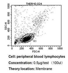
- Experimental details
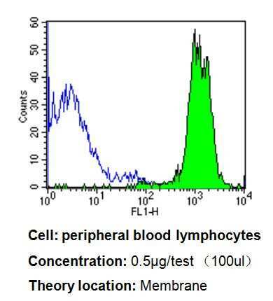
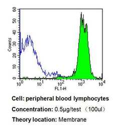
- Flow cytometry analysis of CD45RB in PBMC cells compared to an isotype control (blue). Human blood was collected, combined with a hydrophilic polysaccharide, centrifuged, transferred to a conical tube and washed with PBS. 50 µL of cell solution was added to each tube at a dilution of 2x10^7 cells/mL, followed by the addition of 50 µL of isotype control and primary antibody (Product # MA5-12496) at a dilution of 0.5 µg/test. Cells were incubated for 30 min at 4°C and washed with a cell buffer, followed by incubation with a DyLight 488-conjugated goat anti-mouse IgG (H+L) secondary for 30 min at 4°C in the dark. FACS analysis was performed using 400 µL of cell buffer.
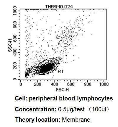
- Submitted by
- Invitrogen Antibodies (provider)
- Main image
- Experimental details
- Flow cytometry analysis of CD45RB in PBMC cells compared to an isotype control (blue). Human blood was collected, combined with a hydrophilic polysaccharide, centrifuged, transferred to a conical tube and washed with PBS. 50 µL of cell solution was added to each tube at a dilution of 2x10^7 cells/mL, followed by the addition of 50 µL of isotype control and primary antibody (Product # MA5-12496) at a dilution of 0.5 µg/test. Cells were incubated for 30 min at 4°C and washed with a cell buffer, followed by incubation with a DyLight 488-conjugated goat anti-mouse IgG (H+L) secondary for 30 min at 4°C in the dark. FACS analysis was performed using 400 µL of cell buffer.