Explore
 Validate
 Learn
Learn Western blot
 Immunocytochemistry
Antibody data
- Antibody Data
- Antigen structure
- References [0]
- Comments [0]
- Validations
- Western blot [1]
Submit
Validation data
Reference
Comment
Report error
- Product number
- P00057 - Provider product page

- Provider
- Boster Biological Technology
- Product name
- Anti-Phospho-ER alpha (S118) ESR1 Rabbit Monoclonal Antibody
- Antibody type
- Monoclonal
- Description
- Monoclonal antibody for ESTROGEN RECEPTOR ALPHA/ESR1 detection. Host: Rabbit.Size: 100ug/vial. Tested applications: IF, IHC, ICC, WB. Reactive species: Human ESTROGEN RECEPTOR ALPHA/ESR1 information: Molecular Weight: 66216 MW; Subcellular Localization: Isoform 1: Nucleus . Cytoplasm . Cell membrane ; Peripheral membrane protein ; Cytoplasmic side . A minor fraction is associated with the inner membrane; Tissue Specificity: Widely expressed. Isoform 3 is not expressed in the pituitary gland.
- Reactivity
- Human
- Host
- Rabbit
- Antibody clone number
- EIG-5
- Vial size
- 100ug/vial
- Concentration
- 0.5-1mg/ml, actual concentration vary by lot. Use suggested dilution ratio to decide dilution procedure.
- Storage
- At -20°C for one year. Avoid repeated freezing and thawing.
No comments: Submit comment
Supportive validation
- Submitted by
- Boster Biological Technology (provider)
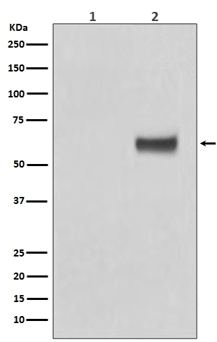
- Main image
- Experimental details
- Western blot analysis of Phospho-ER alpha (S118) expression in (1) MCF7 cell lysate; (2) MCF7 cell lysate treated with b-Estradiol and EGF (P00057). Electrophoresis was performed on a 5-20% SDS-PAGE gel at 70V (Stacking gel) / 90V (Resolving gel) for 2-3 hours. The sample well of each lane was loaded with 50ug of sample under reducing conditions. After Electrophoresis, proteins were transferred to a Nitrocellulose membrane at 150mA for 50-90 minutes. Blocked the membrane with 5% Non-fat Milk/ TBS for 1.5 hour at RT. The membrane was incubated with rabbit anti-ESR1 monoclonal antibody (Catalog # P00057) overnight at 4°C, then washed with TBS-0.1%Tween 3 times with 5 minutes each and probed with a goat anti-rabbit IgG-HRP secondary antibody at a dilution of 1:10000 for 1.5 hour at RT. The signal is developed using an Enhanced Chemiluminescent detection (ECL) kit (Catalog # EK1002) with Tanon 5200 system. A specific band was detected for ESR1
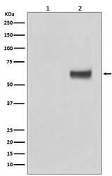
- Additional image