Explore
 Validate
 Learn
Learn Western blot
 Immunocytochemistry
 Immunohistochemistry
Antibody data
- Antibody Data
- Antigen structure
- References [48]
- Comments [0]
- Validations
- Immunocytochemistry [2]
- Other assay [12]
Submit
Validation data
Reference
Comment
Report error
- Product number
- MA1-310 - Provider product page

- Provider
- Invitrogen Antibodies
- Product name
- Estrogen Receptor alpha Monoclonal Antibody (33)
- Antibody type
- Monoclonal
- Antigen
- Synthetic peptide
- Description
- MA1-310 detects both the steroid occupied and unoccupied forms of the estrogen receptor (ER) alpha from human samples. MA1-310 detects ER alpha and does not cross react with ER beta. MA1-310 has been successfully used in Western blot, immunohistochemistry, immunoprecipitation, gel shift, flow cytometry, and immunocytochemical experiments. By Western blot, this antibody detects an ~66 kDa protein representing ER alpha from MCF-7 cell extract. MA1-310 can be used with blocking peptide PEP-013.
- Reactivity
- Human, Mouse, Rat, Bovine
- Host
- Mouse
- Isotype
- IgG
- Antibody clone number
- 33
- Vial size
- 100 μg
- Concentration
- 1 mg/mL
- Storage
- -20°C, Avoid Freeze/Thaw Cycles
Submitted references Immunofluorescent Evidence for Nuclear Localization of Aromatase in Astrocytes in the Rat Central Nervous System.
Regulation of uterine function during estrous cycle, anestrus phase and pregnancy by steroids in red deer (Cervus elaphus L.).
Fetal Zone Steroids and Estrogen Show Sex Specific Effects on Oligodendrocyte Precursor Cells in Response to Oxidative Damage.
Dichotomic effects of clinically used drugs on tumor growth, bone remodeling and pain management.
Estrogen receptor beta signaling inhibits PDGF induced human airway smooth muscle proliferation.
Tibolone protects astrocytic cells from glucose deprivation through a mechanism involving estrogen receptor beta and the upregulation of neuroglobin expression.
Highly variable cancer subpopulations that exhibit enhanced transcriptome variability and metastatic fitness.
Estrogen Enhances Linkage in the Vascular Endothelial Calmodulin Network via a Feedforward Mechanism at the G Protein-coupled Estrogen Receptor 1.
Inhibition of SHP2 in basal-like and triple-negative breast cells induces basal-to-luminal transition, hormone dependency, and sensitivity to anti-hormone treatment.
Increased gene copy number of VAMP7 disrupts human male urogenital development through altered estrogen action.
17β-Estradiol induces supernumerary primordial germ cells in embryos of the polychaete Platynereis dumerilii.
Gender matters: estrogen receptor alpha (ERα) and histone deacetylase (HDAC) 1 and 2 control the gender-specific transcriptional regulation of human uridine diphosphate glucuronosyltransferases genes (UGT1A).
Characterization of estrogen response element binding proteins as biomarkers of breast cancer behavior.
Effect of raloxifene and atorvastatin in atherosclerotic process in ovariectomized rats.
Binding of the ERα and ARNT1 AF2 domains to exon 21 of the SRC1 isoform SRC1e is essential for estrogen- and dioxin-related transcription.
Estradiol inhibits chondrogenic differentiation of mesenchymal stem cells via nonclassic signaling.
Developmental programming: prenatal androgen excess disrupts ovarian steroid receptor balance.
Modulation of androgen and estrogen receptor expression by antiepileptic drugs and steroids in hippocampus of patients with temporal lobe epilepsy.
Estrous cycle-dependent expression of estrogen receptor alpha in periodontal tissue.
Quantitative determination of steroid hormone receptor positive cells in the synovium of patients with rheumatoid arthritis and osteoarthritis: is there a link to inflammation?
Sex steroid receptor evolution and signalling in aquatic invertebrates.
A novel role for the glucocorticoid receptor in the regulation of monocyte chemoattractant protein-1 mRNA stability.
Expression of the estrogen receptor in blood neutrophils of dairy cows during the periparturient period.
Expression of estrogen receptor alpha and 17beta-hydroxysteroid dehydrogenase 4 in the ciliary body.
Effects of cadmium on the reproductive axis of Japanese medaka (Oryzias latipes).
Effects of cadmium on the reproductive axis of Japanese medaka (Oryzias latipes).
Effects of an antiprogestin onapristone on the endometrium of bonnet monkeys: morphometric and ultrastructural studies.
Expression of estrogen receptor alpha and beta in the epiphyseal plate of the rat.
Linkage of rapid estrogen action to MAPK activation by ERalpha-Shc association and Shc pathway activation.
Osteoblast differentiation influences androgen and estrogen receptor-alpha and -beta expression.
Osteoblast differentiation influences androgen and estrogen receptor-alpha and -beta expression.
Transgenerational and developmental exposure of Japanese medaka (Oryzias latipes) to ethinylestradiol results in endocrine and reproductive differences in the response to ethinylestradiol as adults.
Transgenerational and developmental exposure of Japanese medaka (Oryzias latipes) to ethinylestradiol results in endocrine and reproductive differences in the response to ethinylestradiol as adults.
Expression of estrogen receptor in the choroidal neovascular membranes in highly myopic eyes.
Measurement of estrogen receptors in intact cells by flow cytometry.
Evidence for estrogen receptor expression in germ cell and somatic cell subpopulations in the ovary of the newly hatched chicken.
17 Beta-estradiol increases VEGF receptor-2 and promotes DNA synthesis in retinal microvascular endothelial cells.
The negative regulation of the rat aldehyde dehydrogenase 3 gene by glucocorticoids: involvement of a single imperfect palindromic glucocorticoid responsive element.
Estrogen receptor expression in bovine and rat retinas.
Identification of an estrogen response element activated by metabolites of 17beta-estradiol and raloxifene.
Identification of an estrogen response element activated by metabolites of 17beta-estradiol and raloxifene.
Estrogen modulates the recruitment of myelopoietic cell progenitors in rat through a stromal cell-independent mechanism involving apoptosis.
Immunogold labelling of estradiol receptor in MCF7 cells.
Immunogold labelling of estradiol receptor in MCF7 cells.
Immunohistochemical detection and northern blot analysis of estrogen receptor in osteoblastic cells.
Vascular smooth muscle cells as target for estrogen.
Establishment of the human BSMZ breast cancer cell line, which overexpresses the erbB-2 and c-myc genes.
Development and characterization of monoclonal antibodies to a specific domain of human estrogen receptor.
Kata D, Gróf I, Hoyk Z, Ducza E, Deli MA, Zupkó I, Földesi I
International journal of molecular sciences 2022 Aug 11;23(16)
International journal of molecular sciences 2022 Aug 11;23(16)
Regulation of uterine function during estrous cycle, anestrus phase and pregnancy by steroids in red deer (Cervus elaphus L.).
Kotlarczyk AM, Grzyb M, Korzekwa AJ
Scientific reports 2021 Oct 11;11(1):20109
Scientific reports 2021 Oct 11;11(1):20109
Fetal Zone Steroids and Estrogen Show Sex Specific Effects on Oligodendrocyte Precursor Cells in Response to Oxidative Damage.
Sunny DE, Hammer E, Ittermann T, Krüger EL, Hübner S, Hartmann MF, Wudy SA, Völker U, Heckmann M
International journal of molecular sciences 2021 Jun 19;22(12)
International journal of molecular sciences 2021 Jun 19;22(12)
Dichotomic effects of clinically used drugs on tumor growth, bone remodeling and pain management.
Barrière DA, Midavaine É, Doré-Savard L, Kirby K, Tremblay L, Beaudoin JF, Beaudet N, Longpré JM, Lecomte R, Lepage M, Sarret P
Scientific reports 2019 Dec 27;9(1):20155
Scientific reports 2019 Dec 27;9(1):20155
Estrogen receptor beta signaling inhibits PDGF induced human airway smooth muscle proliferation.
Ambhore NS, Katragadda R, Raju Kalidhindi RS, Thompson MA, Pabelick CM, Prakash YS, Sathish V
Molecular and cellular endocrinology 2018 Nov 15;476:37-47
Molecular and cellular endocrinology 2018 Nov 15;476:37-47
Tibolone protects astrocytic cells from glucose deprivation through a mechanism involving estrogen receptor beta and the upregulation of neuroglobin expression.
Avila-Rodriguez M, Garcia-Segura LM, Hidalgo-Lanussa O, Baez E, Gonzalez J, Barreto GE
Molecular and cellular endocrinology 2016 Sep 15;433:35-46
Molecular and cellular endocrinology 2016 Sep 15;433:35-46
Highly variable cancer subpopulations that exhibit enhanced transcriptome variability and metastatic fitness.
Nguyen A, Yoshida M, Goodarzi H, Tavazoie SF
Nature communications 2016 May 3;7:11246
Nature communications 2016 May 3;7:11246
Estrogen Enhances Linkage in the Vascular Endothelial Calmodulin Network via a Feedforward Mechanism at the G Protein-coupled Estrogen Receptor 1.
Tran QK, Firkins R, Giles J, Francis S, Matnishian V, Tran P, VerMeer M, Jasurda J, Burgard MA, Gebert-Oberle B
The Journal of biological chemistry 2016 May 13;291(20):10805-23
The Journal of biological chemistry 2016 May 13;291(20):10805-23
Inhibition of SHP2 in basal-like and triple-negative breast cells induces basal-to-luminal transition, hormone dependency, and sensitivity to anti-hormone treatment.
Zhao H, Agazie YM
BMC cancer 2015 Mar 8;15:109
BMC cancer 2015 Mar 8;15:109
Increased gene copy number of VAMP7 disrupts human male urogenital development through altered estrogen action.
Tannour-Louet M, Han S, Louet JF, Zhang B, Romero K, Addai J, Sahin A, Cheung SW, Lamb DJ
Nature medicine 2014 Jul;20(7):715-24
Nature medicine 2014 Jul;20(7):715-24
17β-Estradiol induces supernumerary primordial germ cells in embryos of the polychaete Platynereis dumerilii.
Lidke AK, Bannister S, Löwer AM, Apel DM, Podleschny M, Kollmann M, Ackermann CF, García-Alonso J, Raible F, Rebscher N
General and comparative endocrinology 2014 Jan 15;196:52-61
General and comparative endocrinology 2014 Jan 15;196:52-61
Gender matters: estrogen receptor alpha (ERα) and histone deacetylase (HDAC) 1 and 2 control the gender-specific transcriptional regulation of human uridine diphosphate glucuronosyltransferases genes (UGT1A).
Kalthoff S, Winkler A, Freiberg N, Manns MP, Strassburg CP
Journal of hepatology 2013 Oct;59(4):797-804
Journal of hepatology 2013 Oct;59(4):797-804
Characterization of estrogen response element binding proteins as biomarkers of breast cancer behavior.
Kruer TL, Cummins TD, Powell DW, Wittliff JL
Clinical biochemistry 2013 Nov;46(16-17):1739-46
Clinical biochemistry 2013 Nov;46(16-17):1739-46
Effect of raloxifene and atorvastatin in atherosclerotic process in ovariectomized rats.
Cetinkaya Demir B, Uyar Y, Ozbilgin K, Köse C
The journal of obstetrics and gynaecology research 2013 Jan;39(1):229-36
The journal of obstetrics and gynaecology research 2013 Jan;39(1):229-36
Binding of the ERα and ARNT1 AF2 domains to exon 21 of the SRC1 isoform SRC1e is essential for estrogen- and dioxin-related transcription.
Endler A, Chen L, Zhang J, Xu GT, Shibasaki F
Journal of cell science 2012 Apr 15;125(Pt 8):2004-16
Journal of cell science 2012 Apr 15;125(Pt 8):2004-16
Estradiol inhibits chondrogenic differentiation of mesenchymal stem cells via nonclassic signaling.
Jenei-Lanzl Z, Straub RH, Dienstknecht T, Huber M, Hager M, Grässel S, Kujat R, Angele MK, Nerlich M, Angele P
Arthritis and rheumatism 2010 Apr;62(4):1088-96
Arthritis and rheumatism 2010 Apr;62(4):1088-96
Developmental programming: prenatal androgen excess disrupts ovarian steroid receptor balance.
Ortega HH, Salvetti NR, Padmanabhan V
Reproduction (Cambridge, England) 2009 May;137(5):865-77
Reproduction (Cambridge, England) 2009 May;137(5):865-77
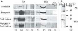
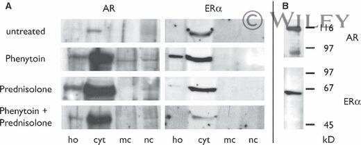
Modulation of androgen and estrogen receptor expression by antiepileptic drugs and steroids in hippocampus of patients with temporal lobe epilepsy.
Killer N, Hock M, Gehlhaus M, Capetian P, Knoth R, Pantazis G, Volk B, Meyer RP
Epilepsia 2009 Aug;50(8):1875-90
Epilepsia 2009 Aug;50(8):1875-90
Estrous cycle-dependent expression of estrogen receptor alpha in periodontal tissue.
Zhao Q, Tan Z, Guo J, Chen Y
Chronobiology international 2007;24(3):425-33
Chronobiology international 2007;24(3):425-33
Quantitative determination of steroid hormone receptor positive cells in the synovium of patients with rheumatoid arthritis and osteoarthritis: is there a link to inflammation?
Capellino S, Riepl B, Rauch L, Angele P, Cutolo M, Straub RH
Annals of the rheumatic diseases 2007 Jan;66(1):53-8
Annals of the rheumatic diseases 2007 Jan;66(1):53-8
Sex steroid receptor evolution and signalling in aquatic invertebrates.
Köhler HR, Kloas W, Schirling M, Lutz I, Reye AL, Langen JS, Triebskorn R, Nagel R, Schönfelder G
Ecotoxicology (London, England) 2007 Feb;16(1):131-43
Ecotoxicology (London, England) 2007 Feb;16(1):131-43
A novel role for the glucocorticoid receptor in the regulation of monocyte chemoattractant protein-1 mRNA stability.
Dhawan L, Liu B, Blaxall BC, Taubman MB
The Journal of biological chemistry 2007 Apr 6;282(14):10146-52
The Journal of biological chemistry 2007 Apr 6;282(14):10146-52
Expression of the estrogen receptor in blood neutrophils of dairy cows during the periparturient period.
Lamote I, Meyer E, De Ketelaere A, Duchateau L, Burvenich C
Theriogenology 2006 Apr 1;65(6):1082-98
Theriogenology 2006 Apr 1;65(6):1082-98
Expression of estrogen receptor alpha and 17beta-hydroxysteroid dehydrogenase 4 in the ciliary body.
Kobayashi K, Iwakiri R, Kobayashi H, Okinami S
Graefe's archive for clinical and experimental ophthalmology = Albrecht von Graefes Archiv fur klinische und experimentelle Ophthalmologie 2004 Feb;242(2):172-176
Graefe's archive for clinical and experimental ophthalmology = Albrecht von Graefes Archiv fur klinische und experimentelle Ophthalmologie 2004 Feb;242(2):172-176
Effects of cadmium on the reproductive axis of Japanese medaka (Oryzias latipes).
Tilton SC, Foran CM, Benson WH
Comparative biochemistry and physiology. Toxicology & pharmacology : CBP 2003 Nov;136(3):265-76
Comparative biochemistry and physiology. Toxicology & pharmacology : CBP 2003 Nov;136(3):265-76
Effects of an antiprogestin onapristone on the endometrium of bonnet monkeys: morphometric and ultrastructural studies.
Gopalkrishnan K, Katkam RR, Sachdeva G, Kholkute SD, Padwal V, Puri CP
Biology of reproduction 2003 Jun;68(6):1959-67
Biology of reproduction 2003 Jun;68(6):1959-67
Expression of estrogen receptor alpha and beta in the epiphyseal plate of the rat.
van der Eerden BC, Gevers EF, Löwik CW, Karperien M, Wit JM
Bone 2002 Mar;30(3):478-85
Bone 2002 Mar;30(3):478-85
Linkage of rapid estrogen action to MAPK activation by ERalpha-Shc association and Shc pathway activation.
Song RX, McPherson RA, Adam L, Bao Y, Shupnik M, Kumar R, Santen RJ
Molecular endocrinology (Baltimore, Md.) 2002 Jan;16(1):116-27
Molecular endocrinology (Baltimore, Md.) 2002 Jan;16(1):116-27
Osteoblast differentiation influences androgen and estrogen receptor-alpha and -beta expression.
Wiren KM, Chapman Evans A, Zhang XW
The Journal of endocrinology 2002 Dec;175(3):683-94
The Journal of endocrinology 2002 Dec;175(3):683-94
Osteoblast differentiation influences androgen and estrogen receptor-alpha and -beta expression.
Wiren KM, Chapman Evans A, Zhang XW
The Journal of endocrinology 2002 Dec;175(3):683-94
The Journal of endocrinology 2002 Dec;175(3):683-94
Transgenerational and developmental exposure of Japanese medaka (Oryzias latipes) to ethinylestradiol results in endocrine and reproductive differences in the response to ethinylestradiol as adults.
Foran CM, Peterson BN, Benson WH
Toxicological sciences : an official journal of the Society of Toxicology 2002 Aug;68(2):389-402
Toxicological sciences : an official journal of the Society of Toxicology 2002 Aug;68(2):389-402
Expression of estrogen receptor in the choroidal neovascular membranes in highly myopic eyes.
Kobayashi K, Mandai M, Suzuma I, Kobayashi H, Okinami S
Retina (Philadelphia, Pa.) 2002 Aug;22(4):418-22
Retina (Philadelphia, Pa.) 2002 Aug;22(4):418-22
Measurement of estrogen receptors in intact cells by flow cytometry.
Cao S, Hudnall SD, Kohen F, Lu LJ
Cytometry 2000 Oct 1;41(2):109-14
Cytometry 2000 Oct 1;41(2):109-14
Evidence for estrogen receptor expression in germ cell and somatic cell subpopulations in the ovary of the newly hatched chicken.
Méndez MC, Chávez B, Echeverría O, Vilchis F, Vázquez Nin GH, Pedernera E
Cell and tissue research 1999 Oct;298(1):145-52
Cell and tissue research 1999 Oct;298(1):145-52
17 Beta-estradiol increases VEGF receptor-2 and promotes DNA synthesis in retinal microvascular endothelial cells.
Suzuma I, Mandai M, Takagi H, Suzuma K, Otani A, Oh H, Kobayashi K, Honda Y
Investigative ophthalmology & visual science 1999 Aug;40(9):2122-9
Investigative ophthalmology & visual science 1999 Aug;40(9):2122-9
The negative regulation of the rat aldehyde dehydrogenase 3 gene by glucocorticoids: involvement of a single imperfect palindromic glucocorticoid responsive element.
Falkner KC, Xiao GH, Pinaire JA, Pendleton ML, Lindahl R, Prough RA
Molecular pharmacology 1999 Apr;55(4):649-57
Molecular pharmacology 1999 Apr;55(4):649-57
Estrogen receptor expression in bovine and rat retinas.
Kobayashi K, Kobayashi H, Ueda M, Honda Y
Investigative ophthalmology & visual science 1998 Oct;39(11):2105-10
Investigative ophthalmology & visual science 1998 Oct;39(11):2105-10
Identification of an estrogen response element activated by metabolites of 17beta-estradiol and raloxifene.
Yang NN, Venugopalan M, Hardikar S, Glasebrook A
Science (New York, N.Y.) 1996 Aug 30;273(5279):1222-5
Science (New York, N.Y.) 1996 Aug 30;273(5279):1222-5
Identification of an estrogen response element activated by metabolites of 17beta-estradiol and raloxifene.
Yang NN, Venugopalan M, Hardikar S, Glasebrook A
Science (New York, N.Y.) 1996 Aug 30;273(5279):1222-5
Science (New York, N.Y.) 1996 Aug 30;273(5279):1222-5
Estrogen modulates the recruitment of myelopoietic cell progenitors in rat through a stromal cell-independent mechanism involving apoptosis.
Shevde NK, Pike JW
Blood 1996 Apr 1;87(7):2683-92
Blood 1996 Apr 1;87(7):2683-92
Immunogold labelling of estradiol receptor in MCF7 cells.
Sierralta WD, Bönig I, Thole HH
Cell and tissue research 1995 Mar;279(3):445-52
Cell and tissue research 1995 Mar;279(3):445-52
Immunogold labelling of estradiol receptor in MCF7 cells.
Sierralta WD, Bönig I, Thole HH
Cell and tissue research 1995 Mar;279(3):445-52
Cell and tissue research 1995 Mar;279(3):445-52
Immunohistochemical detection and northern blot analysis of estrogen receptor in osteoblastic cells.
Ikegami A, Inoue S, Hosoi T, Mizuno Y, Nakamura T, Ouchi Y, Orimo H
Journal of bone and mineral research : the official journal of the American Society for Bone and Mineral Research 1993 Sep;8(9):1103-9
Journal of bone and mineral research : the official journal of the American Society for Bone and Mineral Research 1993 Sep;8(9):1103-9
Vascular smooth muscle cells as target for estrogen.
Orimo A, Inoue S, Ikegami A, Hosoi T, Akishita M, Ouchi Y, Muramatsu M, Orimo H
Biochemical and biophysical research communications 1993 Sep 15;195(2):730-6
Biochemical and biophysical research communications 1993 Sep 15;195(2):730-6
Establishment of the human BSMZ breast cancer cell line, which overexpresses the erbB-2 and c-myc genes.
Watanabe M, Tanaka H, Kamada M, Okano JH, Takahashi H, Uchida K, Iwamura A, Zeniya M, Ohno T
Cancer research 1992 Oct 1;52(19):5178-82
Cancer research 1992 Oct 1;52(19):5178-82
Development and characterization of monoclonal antibodies to a specific domain of human estrogen receptor.
Traish AM, Ettinger R, Kim N, Marshak-Rothstein A, Wotiz HH
Steroids 1990 May;55(5):196-208
Steroids 1990 May;55(5):196-208
No comments: Submit comment
Supportive validation
- Submitted by
- Invitrogen Antibodies (provider)
- Main image
- Experimental details
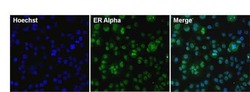
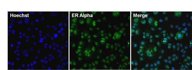
- Immunofluorescent analysis of Estrogen Receptor Alpha (green) in T47D cells. The cells were fixed with formalin for 15 minutes, permeabilized with 0.1% Triton X-100 in TBS for 10 minutes, and blocked with 3% BSA (Product # 37525) for 30 minutes at room temperature. Cells were stained with or without Estrogen Receptor Alpha monoclonal antibody (Product # MA1-310), at a concentration of 10 µg/mL overnight at 4C, and then incubated with a Superclonal goat anti-mouse IgG Alexa Fluor 488 secondary antibody (Product # A28175) at a dilution of 1:1000 for 30 minutes at room temperature (both panels, green). Nuclei (both panels, blue) were stained with Hoechst 33342 dye (Product # 62249). Images were taken on a Thermo Scientific ToxInsight at 20X magnification.
- Submitted by
- Invitrogen Antibodies (provider)
- Main image

- Experimental details
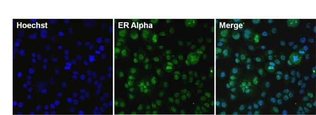
- Immunofluorescent analysis of Estrogen Receptor Alpha (green) in T47D cells. The cells were fixed with formalin for 15 minutes, permeabilized with 0.1% Triton X-100 in TBS for 10 minutes, and blocked with 3% BSA (Product # 37525) for 30 minutes at room temperature. Cells were stained with or without Estrogen Receptor Alpha monoclonal antibody (Product # MA1-310), at a concentration of 10 µg/mL overnight at 4C, and then incubated with a Superclonal goat anti-mouse IgG Alexa Fluor 488 secondary antibody (Product # A28175) at a dilution of 1:1000 for 30 minutes at room temperature (both panels, green). Nuclei (both panels, blue) were stained with Hoechst 33342 dye (Product # 62249). Images were taken on a Thermo Scientific ToxInsight at 20X magnification.
Supportive validation
- Submitted by
- Invitrogen Antibodies (provider)
- Main image
- Experimental details
- NULL
- Submitted by
- Invitrogen Antibodies (provider)
- Main image
- Experimental details
- NULL
- Submitted by
- Invitrogen Antibodies (provider)
- Main image
- Experimental details
- NULL

- Submitted by
- Invitrogen Antibodies (provider)
- Main image
- Experimental details
- NULL

- Submitted by
- Invitrogen Antibodies (provider)
- Main image
- Experimental details
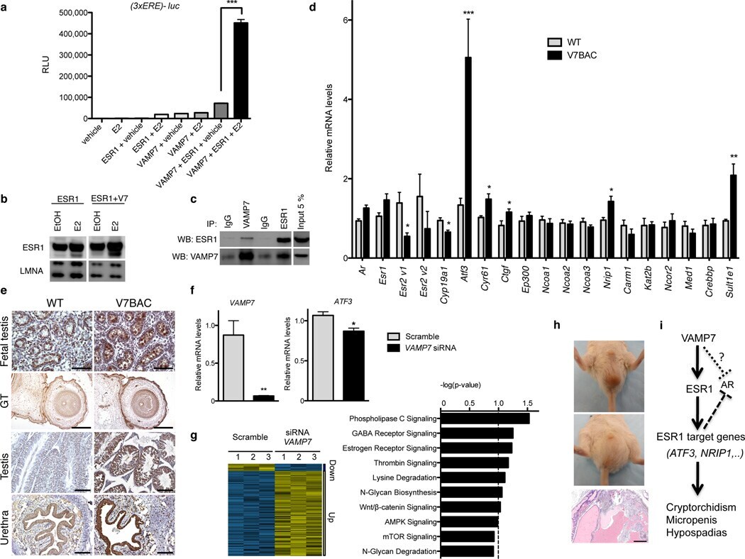
- Figure 5 VAMP7 enhances estrogen receptor transcriptional activity. ( a ) Luciferase assays following transfection with VAMP7 or ESR1 or VAMP7 and ESR1 in HeLa cells incubated in absence (EtOH) or presence of 17 beta-estradiol (10 -8 M) for 24 h. n = 3 independent experiments for each condition. Data are presented as means +- s.e.m. One-way analysis of variance (ANOVA) with post hoc Bonferroni test was used for statistical analyses. * P < 0.05; ** P < 0.01; *** P < 0.001. ( b ) Western Blot analysis of ESR1 and LMNA (lamin A/C) in nuclear protein extracts of HeLa cells co-transfected with ESR1 or ESR1 and VAMP7 in absence (EtOH) or presence of 17 beta-estradiol (10 -8 M) for 24 h. ( c ) Reciprocal co-immunoprecipitation of ESR1 and VAMP7 following their co-transfection in HeLa cells. ( d ) qRT-PCR analysis of key genes of ESR1 signaling in testis from WT ( n = 3) and V7BAC mice (line 7; n = 3). Data are expressed as mean +- s.e.m. Mean differences between WT and V7BAC were determined by unpaired, two-tailed Student's t -test. * P < 0.05; ** P < 0.01; *** P < 0.001. ( e ) ATF3 immunostaining of testis and external genitalia at fetal and adult stages of WT and V7BAC mice. For fetal tissues: scale bar, 250 um. For adult urethra and testis: scale bar, 100 um. ( f ) qRT-PCR of VAMP 7 and ATF3 gene expression after incubation with non-targeting (scramble) or VAMP7 siRNA in NT2/D1. n = 3 independent experiments for each condition. Mean differences were determined by unpaired, t
- Submitted by
- Invitrogen Antibodies (provider)
- Main image

- Experimental details
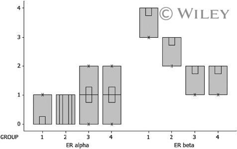
- 1 Estrogen receptor (ER) staining intensities of endothelial cells. Boxplot graphic of ERalpha and ERbeta. Group 1, OVX; Group 2, OVX+RL; Group 3, OVX+AV; Group 4, OVX+RL+AV; Data are presented as median. Kruskal-Wallis test was performed to determine the differences between groups.
- Submitted by
- Invitrogen Antibodies (provider)
- Main image
- Experimental details
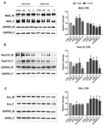
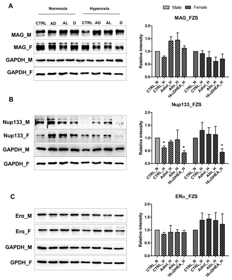
- Figure 3 FZS and E2 show discrete effects on male and female OPCs. Western blot results showing the expression of ( A ) MAG, ( B ) Nup133 and ( C ) ERalpha in OPCs after treatment with 100 nM Adiol (AD), 100 nM Allopregnanolone (AL) and 100 nM 16alpha-OH-DHEA under normoxia and hyperoxia. All data are representative of at least three independent experiments. Bars and error represent mean +- SEM of replicate measurements. Significant differences from normoxia control are indicated by * p < 0.05 (Student's t -test).
- Submitted by
- Invitrogen Antibodies (provider)
- Main image
- Experimental details
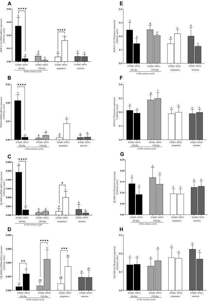
- Figure 1 mRNA and protein expression of AKR1C1 ( A , E ), P450 ( B , F ), 3beta-HSD ( C , G ) and 17beta-HSD ( D , H ) in uterine tissues (endometrium and myometrium) on 4th and 13th day of estrous cycle, in pregnancy and anestrus phase. Data were normalized against GAPDH for mRNA expression and against beta-actin (ACTB) for proteins expression. Each bar represents one experimental group with SEM. Statistical differences were analyzed by two-way analysis (ANOVA) of variance followed by the Bonferroni post hoc test using GraphPad PRISM (Version 8.3.0). The lowest statistical significance was P < 0.05. Asterisks indicate statistical differences between endometrium and myometrium (* P < 0.1; ** P < 0.01; *** P < 0.001; **** P < 0.0001). Different letters indicate statistical differences ( P < 0.05) between the experimental groups throughout endometrium (A, B) and myometrium (a-b) respectively.
- Submitted by
- Invitrogen Antibodies (provider)
- Main image

- Experimental details
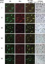
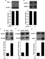
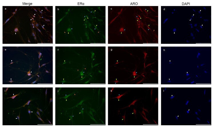
- Nuclear aromatase expression overlaps with nuclear ERalpha signal. Double immunostaining with ERalpha (green) and Aro (red) in glia culture ( a - l ) shows that nuclear Aro signals colocalize with nuclear ERalpha ( a , e , i ; white arrowheads). Both Aro and ERalpha are strongly represented in the nucleus ( b , c , f , g , j,k ; yellow arrowheads) and also in the cytoplasm ( b , c , f , g , j , k ; blue arrowheads). ERalpha positive dots are also detectable in the processes ( b , f,j ; orange arrowheads). Scale bar: 100 mum.
- Submitted by
- Invitrogen Antibodies (provider)
- Main image

- Experimental details
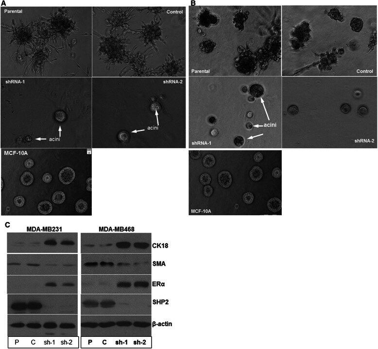
- Figure 4 Silencing SHP2 expression induces acini-like structure formation. A) Light pictures of parental, control and SHP2-silenced MDA-MB231 cells cultured in 3D LRBM matrigel that allows acini-like structure formation. B) Light pictures of parental, control and SHP2-silenced MDA-MB468 cells cultured in 3D LRBM matrigel that allows acini-like structure formation. Pictures of acini-like structures formed by the MCF-10A cells cultured under identical conditions is shown in both A and B for comparison. C) Effect of SHP2 silencing on expression of basal and luminal markers and estrogen receptor alpha (ERalpha). Data presented is representative of at least three independent experiments.
- Submitted by
- Invitrogen Antibodies (provider)
- Main image
- Experimental details
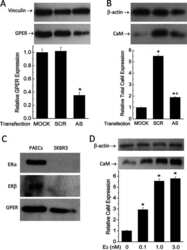
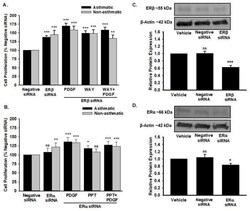
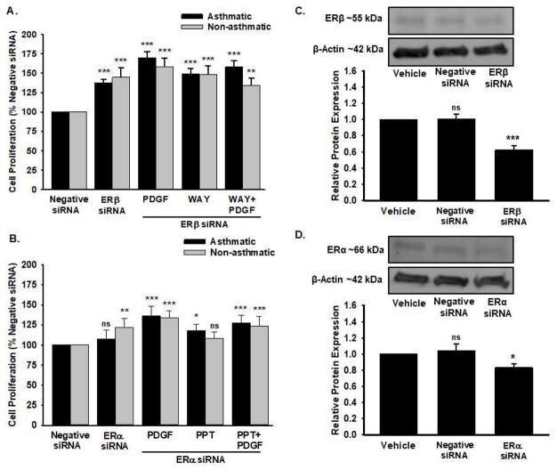
- Effect of ER siRNA on human ASM proliferation and ER Protein Expression MTT assay was used to study the effect of ERβ agonist WAY and ER&alpha agonist PPT on PDGF-induced proliferation in Asthmatic and Non-Asthmatic human ASM cells transfected with ERβ siRNA (Figure 5A) and ERα siRNA (Figure 5B) respectively. There was significant mitogenic effect observed in ERβ siRNA transfected cells compared to negative siRNA in both asthmatic and non-asthmatic ASM cells. ERβ siRNA transfected human ASM cells treated with WAY and WAY+PDGF produced a significant increase in proliferation as compared to negative siRNA treated group. PPT+PDGF treatment produced an increased proliferation in ERα siRNA transfected human ASM cells. Western blot analysis was performed to determine the transfection efficacy of ERβ (Figure 5C) and ERα (Figure 5D) receptor siRNA in human ASM cells. Values are means SEM from n of 5 samples from asthmatics and n of 6 samples from non-asthmatics for MTT assay. Values are means SEM from n of 3 different patient samples of asthmatics and non-asthmatics for western analysis. ***p
- Submitted by
- Invitrogen Antibodies (provider)
- Main image
- Experimental details
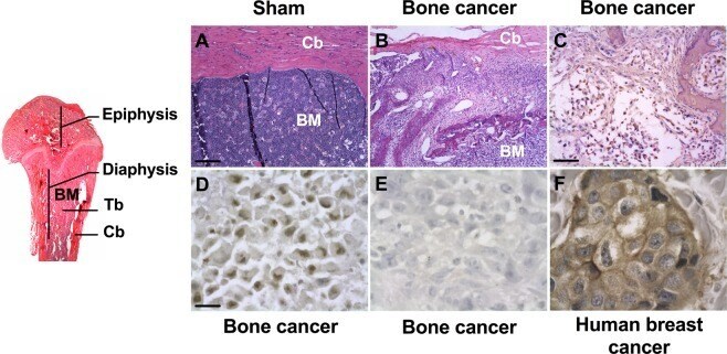
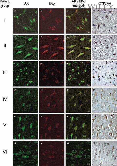
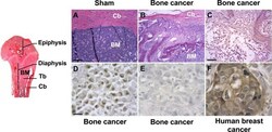
- Figure 1 Histopathological and molecular characteristics of bone metastases in breast cancer, following intra-femoral inoculation of syngeneic MRMT-1 tumor cells. ( A ) Sham-operated female rats display a healthy femoral bone marrow (BM) and cortical bone (Cb) while ( B ) tumor-bearing rats exhibit intense cancer cell invasion into the bone marrow, 18 days following cancer cell implantation. ( C ) Extensive Ki-67-positive staining (brown-stained cancer cell nuclei) indicates a highly proliferative tumor, also expressing ERalpha (estrogen alpha receptor) ( D ) but not the HER-2 receptor ( E ). An anonymous human HER-2 breast cancer sample is used here as a positive control for HER-2 staining ( F ). Scale bars represent 100 um in ( A , B ), 200 um in ( C ) and 50 um in ( D-F ).